Clipart tagged: ‘Choroid’
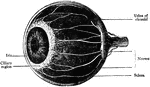

Eye
"Next in order is the aqueous humor, b, e, in the middle of which is the iris, d, c. Behind the pupil…
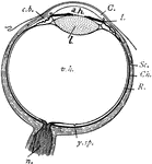
Eye
"Diagram of the eye. C., Cornea; a.h., aqueous humour; c.b., ciliary body; l., lens; I., iris; Sc.,…
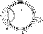
Diagram of the Eye
Plan of the eye seen in section. Labels: A, The Sclerotic Coat; B, The Choroid Coat; C, The Retina;…
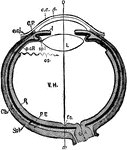
Human Eye
"Diagrammatic horizontal section of the eye of man. c, cornea; ch. choroid (dotted); C. P, ciliary processes;…
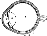
The Eye
The eye. Labels: a, sclerotica; e, cornea; b, choroid; d, optic nerve; f, aqueous humor; g g , iris;…
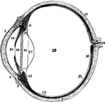
Left Eyeball in Horizontal Section
The left eyeball in horizontal section from before back. Labels: 1, sclerotic; 2, junction of sclerotic…
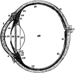
Section of Left Eyeball
The left eyeball in horizontal section from before back. Labels: 1, sclerotic; 2, junction of sclerotic…
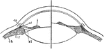
Focusing of the Eye
Diagram to illustrate the mechanism of accommodation (focusing); on the right half of the figure for…
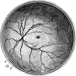
Posterior Half of the Retina
The posterior half of the retina of the left eye, viewed from before; s, the cut edge of the sclerotic…
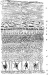
Structure of the Retina
A section of the retina, choroid, and part of the sclerotic. Labels: a, membrana limitans interna; b,…