Clipart tagged: ‘cochlea’
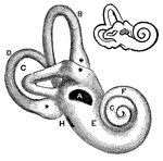
Cat Ear
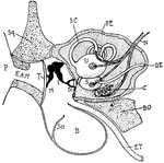
"Diagram showing the ear and related parts in a young cat. P., Pinna; Sq., squamosal: E.A.M., external…
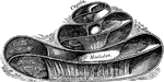
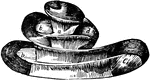
Cochlea
Diagram of a section of a coil of the cochlea of the ear. Labels: C.C, canal of the cochlea; mR, its…
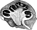
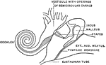
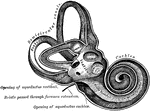
The Cochlea and Passages of the Ear
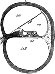
In this figure are shown the winding passages of the ear (the labyrinth of the ear). The middle part…
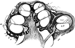
Cochlea in Transverse Section
The cochlea in transverse section. Observe especially the canals of the cochlea, which is a part of…
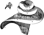
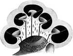
The Cochlea of the Ear
A vertical section of the cochlea, highly magnified, to show the arrangement and connection of its parts.
Longitudinal Section of the Cochlea
Longitudinal section of the cochlea, showing the relations of the scalae, the ganglion spirale, ect.…
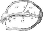
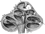
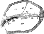
Cochlea, Section of
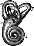
Section of one coil of the cochlea, magnified. Labels: SV, scala vestibuli; R, membrane of Reissner;…
Section Through Coil of the Cochlea
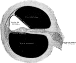
Section through one of the coils of the cochlea. Labels: ST, scala tympani; SV, scala vestibuli; CC,…
The Cochlea
The cochlea of the ear which is a spiral canal situated in the eburnated portion of the petrous bone,…
Vertical Section Through the Cochlea
Vertical section through the right cochlea, medial portion, viewed from the lateral side.
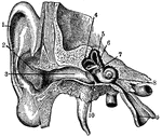
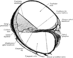
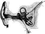
Ear
"Section through right ear. 1, helix; 2, concha; 3, outer passage; 4, 5, 6, semi-circular canals; 7,…
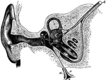
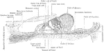
Ear and Auditory Canal
Semi-diagrammatic section through the right ear. Labels: M, concha; G, the external auditory canal;…
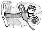
Human Ear
A diagram of the human ear. It is divided into the outer ear - A, middle ear - B, and inner ear - C.…
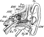
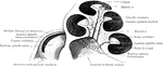
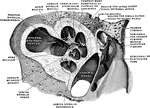
Inner Ear
"Transverse Section through Side Walls of Skull, showing the Inner Parts of the Ear. Co, concha or external…
Section Through the Right Ear
Semi-diagrammatic section through the right ear. Labels: M, concha; G, external auditory meatus; T,…
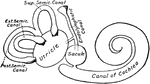
Osseous Labyrinth in Vertical Section
The osseous labyrinth in vertical section. The broken, white lines indicate the position of the basilar…