The cricothyroid muscle.

Cricothyroid Muscle

The cricothyroid muscle.

Diagram to show the course of the esophagus.

Course of the Esophagus

Diagram to show the course of the esophagus.

Muscles of the larynx. Side view. Right ala of thyroid cartilage removed.

Side View of the Muscles of the Larynx

Muscles of the larynx. Side view. Right ala of thyroid cartilage removed.
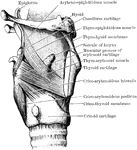
Dissection of the muscles in the lateral wall of the larynx. The right ala of the thyroid cartilage has been removed.

Larynx Muscle

Dissection of the muscles in the lateral wall of the larynx. The right ala of the thyroid cartilage…
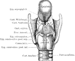
The larynx seen from behind after the removal of the muscles. The cartilages and ligaments only remain.

Larynx Seen from Behind

The larynx seen from behind after the removal of the muscles. The cartilages and ligaments only remain.

The larynx seen from behind to show the intrinsic muscle posteriorly.

Larynx Seen from Behind

The larynx seen from behind to show the intrinsic muscle posteriorly.

Larynx, viewed from above.

Larynx Viewed from Above

Larynx, viewed from above.

Muscles of the larynx, back view.

Back View of the Muscles of the Larynx

Muscles of the larynx, back view.

Coronal section of the larynx, rear view of front half.

Coronal Section of the Larynx

Coronal section of the larynx, rear view of front half.

Coronal section through larynx, to show its three compartments.

Coronal Section Through Larynx

Coronal section through larynx, to show its three compartments.

Cricoid cartilage of the larynx, situated below the thyroid.

Cricoid Cartilage of the Larynx

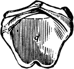
Cricoid cartilage of the larynx, situated below the thyroid.

"Cartilages and Ligaments of the Larynx. (Front view.) <em>A</em>, hyoid bone; <em>B</em>, membrane attached to hyoid bone and the shield-shaped cartilage below (thyroid); edges of this shield-shaped cartilage unite at <em>C</em> (Adam's apple is the V-shaped groove on a line with <em>B</em> and <em>C</em>; <em>D</em>, membrane between the shield-shaped cartilage and the signet-ring cartilage below; <em>E</em> cricoid, or signet-ring, cartilage; <em>F</em>, upper ring of the windpipe." &mdash; Blaisedell, 1904

Front view of the larynx

"Cartilages and Ligaments of the Larynx. (Front view.) A, hyoid bone; B, membrane…
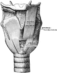
Muscles of the larynx, front view. The Sternothyroid and right Thyrohyoid have been removed.

Front View of the Muscles of the Larynx

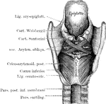
Muscles of the larynx, front view. The Sternothyroid and right Thyrohyoid have been removed.

Interior of the larynx, seen from above.

Interior of the Larynx

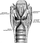
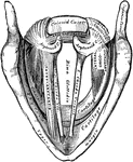
Interior of the larynx, seen from above.

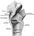
Mesial section through larynx, to show the outer wall of the right half.

Mesial Section Through Larynx

Mesial section through larynx, to show the outer wall of the right half.

"Cartilages and Ligaments of the Larynx. (Front view.) <em>A</em>, epiglottis; <em>B</em>, thyroid cartilage; <em>C</em>, ladle-shaped cartilage; <em>E</em>, cricoid cartilage; <em>F</em>, upper ring of the windpipe." &mdash; Blaisedell, 1904

Posterior view of the larynx

"Cartilages and Ligaments of the Larynx. (Front view.) A, epiglottis; B, thyroid cartilage;…
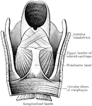
Sagittal section of the larynx, right half.

Sagittal Section of the Larynx

Sagittal section of the larynx, right half.

The lower part of the pharynx and the upper part of the esophagus have been slit up from behind, and the mucous membrane removed to show the muscular fibers. The two longitudinal bands are seen coming round to the front to be attached by a common tendon to the upper border of the cricoid cartilage.

Pharynx and Esophagus

The lower part of the pharynx and the upper part of the esophagus have been slit up from behind, and…

Outline showing the general form of the larynx, trachea, and bronchi, as seen from behind. Labels: h, the great cornu of the hyoid bone; e,epiglottis; t, superior cornu of the thyroid cartilage ; t1, inferior cornu of the thyroid cartilage; a, points to the back of both the arytenoid cartilages, which are surmounted by the cornicula; c, the middle ridge on the back of the cricoid cartilage; tr, the posterior membranous part of the trachea; b, b1, right and left bronchi.

Back View of Respiratory Apparatus

Outline showing the general form of the larynx, trachea, and bronchi, as seen from behind. Labels: h,…

Outline showing the general form of the larynx, trachea, and bronchi, as seen from front. Labels: h, the great cornu of the hyoid bone; e,epiglottis; t, superior and inferior cornu of the thyroid cartilage; c, middle of the cricoid cartilage; tr, the trachea, showing sixteen cartilaginous rings; b, the right bronchus; b1, the left bronchus.

Front View of Respiratory Apparatus

Outline showing the general form of the larynx, trachea, and bronchi, as seen from front. Labels: h,…
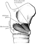
A side view of the thyroid and cricoid cartilages.

Thyroid

A side view of the thyroid and cricoid cartilages.

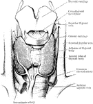
Dissection of the thyroid body and of the parts in immediate relation to it.

Thyroid Body

Dissection of the thyroid body and of the parts in immediate relation to it.

The trachea and bronchi. The thyroid body is indicated by a dotted line.

Trachea and Bronchi

The trachea and bronchi. The thyroid body is indicated by a dotted line.

"<em>A</em>, epiglottis; <em>B</em>, section of hyoid bone; <em>C</em>, false vocal cords which have nothing to do with the production of voice; <em>D</em>, oblong opening between the false and true vocal cords; <em>E</em>, true vocal cord;<em>F</em>, section of the thyroid cartilage; <em>H</em>, section of the anterior portion of the cricoid cartilage; <em>K</em>, trachea; <em>L</em>, section of the posterior portion of the cricoid cartilage; <em>M</em>, ladle-shaped cartilage." &mdash; Blaisedell, 1904

Vocal cords

"A, epiglottis; B, section of hyoid bone; C, false vocal cords which have…