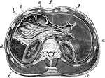
Horizontal section through upper part of abdomen. Labels: a, liver; b, stomach; c, transverse colon; d, spleen; e, kidneys; f, pancreas; g, inferior vena cava; h, aorta with thoracic duct behind it.

Horizontal Section Through Abdomen

Horizontal section through upper part of abdomen. Labels: a, liver; b, stomach; c, transverse colon;…
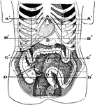
Showing the average position of the abdominal viscera with their surface markings. Labels: A, sterno-ensiform point; A', sterno-ensiform line; B, mid-epigastric point; B', mid-epigastric or transpyloric line; C, umbilical point; C', umbilical line; D, mid-hypogastric point; D', mid-hypogastric line; E, outer border of the right and left rectus abdominis; F, Monro's point- on the right spino-umbilical line at the outer border of the rectus abdomninis.

Abdominal Region

Showing the average position of the abdominal viscera with their surface markings. Labels: A, sterno-ensiform…
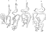
Outlines of the form and position of the alimentary canal in successive stages of its development. A, alimentary canal, in an embryo of four weeks; B, at six weeks; C, at eight weeks; D, at ten weeks; l, the primitive lungs connected with the pharynx; s, the stomach; d, duodenum; i, the small intestine; i', the large intestine; c, the caecum and vermiform appendage; r, the rectum; cl, in A, the cloaca; a, in B, the anus distinct from s i, the sinus uro-genitalis; v, the yolk sac; vi, the vitellointestinal duct; u, the urinary bladder and urachus leading to the allantois; g, genital ducts.

Development of the Alimentary Canal

Outlines of the form and position of the alimentary canal in successive stages of its development. A,…

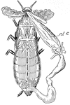
Cockroach dissected to show alimentary canal, al.c.

Cockroach

Cockroach dissected to show alimentary canal, al.c.

Stomach, liver, pancreas, and duodenum of a dog. Labels: a, liver; b, gall bladder; c, biliary canals; d, cystic duct; e, ductus choledocus; f, pancreas; g, pancreatic ducts.

Digestive Organs of a Dog

Stomach, liver, pancreas, and duodenum of a dog. Labels: a, liver; b, gall bladder; c, biliary canals;…
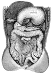
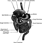
"Showing the Relations of the Stomach, Liver, Intestines, Spleen, and other Organs of the Abdomen. <em>A</em>duodenum</em>; <em>B</em>, upper end of the small intestine; <em>C</em>, lower end of the small intestine; <em>D</em>, caecum; <em>E</em>, bladder. The liver and stomach are drawn up and portions of the lower intesting have been cut away." &mdash; Blaisedell, 1904

Digestive system

"Showing the Relations of the Stomach, Liver, Intestines, Spleen, and other Organs of the Abdomen. Aduodenum;…
Organs of the digestive system.

Organs of the Digestive System

Organs of the digestive system.

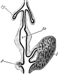
Diagram of part of digestive tract of a chick (4th day). The black line represents hypoblast , the outer shading mesoblast; lg, lung diverticulum with expanded end forming primary lung vesicles; St, stomach; l, two hepatic diverticulum, with their terminations united by solid rows of hypoblast cells; p, diverticulum of the pancreas with the vesicular diverticula coming from it.

Digestive Tract of a Chick

Diagram of part of digestive tract of a chick (4th day). The black line represents hypoblast , the outer…
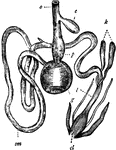
"Digestive system of the common Fowl. o, Gullet; c, Crop; p, Proventriculus; g, Gizzard; sm, Small intestine; k, Intestinal caeca; l, Large intestine; cl, Cloaca." &mdash; Winston's Encyclopedia, 1919

Fowl Digestive System

"Digestive system of the common Fowl. o, Gullet; c, Crop; p, Proventriculus; g, Gizzard; sm, Small intestine;…
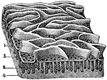
The mucous membrane from the jejunum. Labels: 1, Villi (folds of lining mucous membrane) in miniature. 2, Tubular glands: their orifices. 3, Opening on the free surface of the mucous membrane. 4, Fibrous tissue.

Mucous Membrane from the Jejunum

The mucous membrane from the jejunum. Labels: 1, Villi (folds of lining mucous membrane) in miniature.…

Portion of the mucous membrane from the small intestine, magnified, showing the villi on its free surface, and between them the orifices of the tubular glands. Labels: 1, Portion of a clustered gland. 2, A solitary gland. 3, Fibrous tissue.

A Portion of the Mucous Membrane from the Small Intestine

Portion of the mucous membrane from the small intestine, magnified, showing the villi on its free surface,…
pond mussel dissected to show alimentary canal, al c..

Mussel

pond mussel dissected to show alimentary canal, al c..

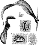
"Pitcher of Nepenthes distillatoria. A, honey-gland from attractive surface of lid; B, digestive gland from interior of pitcher, in pocket-like depression of epidermis, opening downwards; C, transverse section of the same. A, B, and C magnified about 100 diameters." &mdash; The Encyclopedia Britannica, 1893

Nepenthes

"Pitcher of Nepenthes distillatoria. A, honey-gland from attractive surface of lid; B, digestive gland…
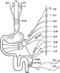
Diagrammatic representation of the nerves of the alimentary canal. Oe to Rct, the various parts of the alimentary canal from esophagus to rectum: L.V, left vagus, ending in front of stomach; rl, recurrent laryngeal nerve, supplying upper part of esophagus; R.V, right vagus, joining left vagus in esophageal plexus; oe.pl., supplying the posterior part of the stomach , and continues at R'V' to join the solar plexus, here represented by a single ganglion, and connected with the inferior mesenteric ganglion m.gl.: a, branches from the solar plexus to stomach and small intestine , and from the mesenteric ganglia and rami communicantes; r.c., belonging to dorsal nerves from the 6th to the 9th (or 10th); Spl.min., small splanchnic nerve similarly from the 10th and 11th dorsal nerves. These both join the solar plexus, and thence make their way to the alimentary canal; c.r., nerves from the ganglia, &c., belonging to 11th and 12th dorsal and 1st and 2nd lumbar nerves, proceeding to the inferior mesenteric ganglia (or plexus), m.gl., and thence by the hypogastic nerve, n.hyp., and the hypogastric nerve, n.hyp., and the hypogastric plexus, pl.hyp., to the circular muscles of the rectum; 1.r., nerves from the 2nd and 3rd sacral nerves, S.2, S.3 (nervi erigentes) proceeding by the hypogastric plexus to the longitudinal muscles of the rectum.

Nerves of the Alimentary Canal

Diagrammatic representation of the nerves of the alimentary canal. Oe to Rct, the various parts of the…
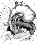
The stomach, pancreas, liver, and duodenum, with part of the rest of the small intestine and the mesentery; the stomach and liver have been turned up so as to expose the pancreas. Labels: V, stomach; D, D',D'', duodenum; L, spleen; P, pancreas; R, right kidney; T, jejunum; Vf, gall bladder; h, hepatic duct; c, cystic duct; ch, common bile duct; 1, aorta; 2, an artery (left coronary) of the stomach; 3, hepatic artery; 4, splenic artery; 5, superior mesenteric artery; 6, superior mesenteric vein; 7, splenic vein; Vp, portal vein.

The Stomach, Pancreas, Liver, and Duodenum

The stomach, pancreas, liver, and duodenum, with part of the rest of the small intestine and the mesentery;…
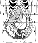
Showing the position of the viscera in the condition of visceroptosis (Glenard's disease). Labels: A, sterno-ensiform line: it crosses above the fifth costal cartilage; B, mid-epigastric line; C, umbilical line; D, mid-hypogastric line; a, pericardium; b, stomach (greatly elongated and dilated); c, liver; c', the lingual or Riedel's process; d, duodenum; e, caecum; f, transverse colon; g, rectum; h, elongated gastro-hepatic omentum.

Position of the Viscera in the Condition of Visceroptosis

Showing the position of the viscera in the condition of visceroptosis (Glenard's disease). Labels: A,…