Diagrammatic transverse section of abdomen, to show the peritoneum on transverse tracing. A, at level of foramen of Winslow. B, lower down. In A note, one of the vasa brevia arteris passing to the stomach between the layers of the gastrosplenic omentum, and also the foramen of Winslow leading into the lesser sac which lies behind the stomach.

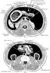
Transverse Section of Abdomen

Diagrammatic transverse section of abdomen, to show the peritoneum on transverse tracing. A, at level…
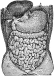
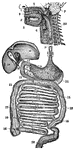
Abdominal organs. Labels: 1, liver turned up; 2, gall bladder; 3, stomach; 4, large intestine; 5, small intestine.

Abdominal Organs

Abdominal organs. Labels: 1, liver turned up; 2, gall bladder; 3, stomach; 4, large intestine; 5, small…
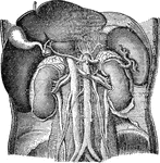
Abdominal organs. Labels: 1, liver turned up; 2, gall bladder; 3, right kidney; 4, spleen; 5, left kidney.

Abdominal Organs

Abdominal organs. Labels: 1, liver turned up; 2, gall bladder; 3, right kidney; 4, spleen; 5, left kidney.

Abdominal organs. Labels: 1, liver; 2, gall bladder; 3, spleen; 4, pancreas.

Abdominal Organs

Abdominal organs. Labels: 1, liver; 2, gall bladder; 3, spleen; 4, pancreas.

Diagram of the abdominal part of the alimentary canal (digestive system). Labels: C, the cardiac, and P, the pyloric end of the stomach; D, the duodenum; J, I, the convolutions of the small intestine; CC, the caecum with the vermiform appendix; AC, ascending, TC, transverse, and DC, descending colon; R, the rectum.

Alimentary Canal

Diagram of the abdominal part of the alimentary canal (digestive system). Labels: C, the cardiac, and…

Front view of two successive stages in the development of the alimentary canal.

Development of the Alimentary Canal

Front view of two successive stages in the development of the alimentary canal.

The digestive apparatus of the horse. Labels: a, mouth; 2, pharynx; 3, esophagus; 4, diaphragm; 5, spleen; 6, stomach (left sac); 7, duodenum; 8, liver (upper extremity); 9, great colon; 10, caecum; 11, small intestine; 12, floating colon; 13, rectum; 14, anus; 15, left kidney and ureter; 16, bladder; 17, urethra; a, hard palate; b, tongue; c, soft palate; d, trachea; e, pulmonary artery (divided); f, heart; g, posterior aorta.

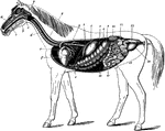
Digestive Apparatus of the Horse

The digestive apparatus of the horse. Labels: a, mouth; 2, pharynx; 3, esophagus; 4, diaphragm; 5, spleen;…

The relation of anterior abdominal digestive organs- left antero-lateral view. Labels: 1, liver; 2, stomach; 3, spleen; 4, colon; a, gastro-hepatic omentum; b, gastro-splenic omentum; c, great omentum; c', its free portion.

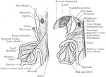
Digestive Organs of a Horse

The relation of anterior abdominal digestive organs- left antero-lateral view. Labels: 1, liver; 2,…
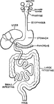
The digestive system.

Digestive System

The digestive system.

A diagram of the organs of digestion. Labels:1, The upper jaw. 2, The lower jaw. 3, The tongue. 4, The roof of the mouth. 5, The esophagus. 6, The trachea. 7, The parotid gland. 8, The sublingual gland. 9, The stomach. 10, The liver. 11, The gall cyst. 12, The duct that conveys the bile to the duodenum (13). 14, The pancreas. 15, The small intestine. 16, The opening of the small intestine into the large intestine. 17, 18, 19, 20, The large intestine. 21, The spleen. 22, The upper part of the spinal column.

Digestive System

A diagram of the organs of digestion. Labels:1, The upper jaw. 2, The lower jaw. 3, The tongue. 4, The…

Two diagrams to illustrate the development of the intestinal canal. The figure to the right shows the rotation of the intestinal loop round the superior mesenteric artery. In both figures the parts are supposed to be viewed from the left side.

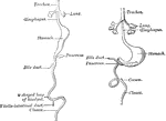
Development of the Intestinal Canal

Two diagrams to illustrate the development of the intestinal canal. The figure to the right shows the…
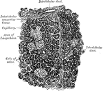
Showing the connection of the small intestine to the thoracic duct by the lacteals lying in the mesentery. Label: Th, thoracic duct.

Lacteals

Showing the connection of the small intestine to the thoracic duct by the lacteals lying in the mesentery.…

Section of human pancreas, showing pancreatic islands.

Section of Pancreas

Section of human pancreas, showing pancreatic islands.

The stomach and intestines. Labels: 1, stomach; 2, duodenum; 3, small intestine; 4, termination of the ileum; 5, caecum; 6, vermiform appendix; 7, ascending colon; 8, transverse colon; 9, descending colon; 10, sigmoid flexure of the colon; 11, rectum; 12, spleen.

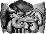
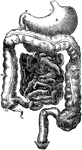
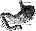
The Stomach and Intestines

The stomach and intestines. Labels: 1, stomach; 2, duodenum; 3, small intestine; 4, termination of the…

The opening part of the stomach where the esophagus joins it is called the cardiac opening; the one at the opposite end, where the small intestine joins it, is called the pylorus.

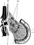
A Section of the Stomach

The opening part of the stomach where the esophagus joins it is called the cardiac opening; the one…

Diagram of the relations of the large intestine and kidneys, from behind.

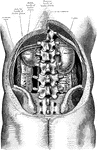
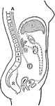
Trunk Showing Organs of Digestion

Diagram of the relations of the large intestine and kidneys, from behind.

Diagram of horizontal section through upper part of 1st lumbar vertebra. The fine dots represent the great sac of the peritoneum, the coarse dots the lesser sac. Labels: A, aorta; Sp, spleen; B.D, bile duct; V.C, vena cava; P, pancreas; H.A, hepatic artery; K, kidney; L, liver; St, stomach; P.V, portal vein.

Horizontal Section Through Trunk

Diagram of horizontal section through upper part of 1st lumbar vertebra. The fine dots represent the…

Transverse section through the middle of the first lumbar vertebra, showing the relations of the pancreas.

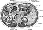
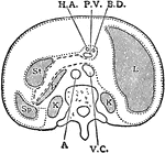
Transverse Section of the Trunk

Transverse section through the middle of the first lumbar vertebra, showing the relations of the pancreas.

Diagram of vertical median section of abdomen. The fine dots represent the great sac of the peritoneum, the coarse dots the lesser sac. Labels: A, aorta; P, pancreas; I, intestine; R, rectum; L, liver; D, duodenum; B, bladder; St, stomach; c, colon; v, vagina.

Vertical Median Section of the Trunk

Diagram of vertical median section of abdomen. The fine dots represent the great sac of the peritoneum,…