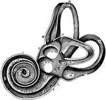
A view of the labyrinth of the left side laid open in its whole extend, so as to show its structure - magnified.

The Labyrinth of the Inner Ear

A view of the labyrinth of the left side laid open in its whole extend, so as to show its structure…
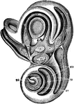
A highly magnified view of the external face of the bony labyrinth of the left side, opened so as to expose the vestibule and its contents, etc.

Bony Labyrinth of the Inner Ear

A highly magnified view of the external face of the bony labyrinth of the left side, opened so as to…
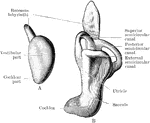
A, Left labyrinth of a human embryo of about four weeks; B, left labyrinth of a human embryo of about 5 weeks.

Development of Labyrinth

A, Left labyrinth of a human embryo of about four weeks; B, left labyrinth of a human embryo of about…
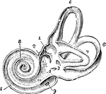
View of the interior of the left labyrinth. The bony wall of the labyrinth is removed superiorly and externally. Labels: 1, fovea hemielliptica; 2m fovea hemispherica; 3, common opening of the superior and posterior semicircular canals; 4, opening of the aqueduct of the vestibule; 5, the superior, 6, the posterior, and 7, the external semicircular canals; 8, spiral tube of the cochlea (scala tympani); 9, opening of the aqueduct of the cochlea; 10, placed on the lamina spiralis in the scala vestibuli.

Interior of the Left Labyrinth

View of the interior of the left labyrinth. The bony wall of the labyrinth is removed superiorly and…
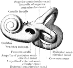
Left bony labyrinth viewed from outer aspect.

Outer Aspect of Labyrinth

Left bony labyrinth viewed from outer aspect.

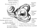
Interior of left bony labyrinth viewed from outer aspect.

Outer Aspect of Interior Labyrinth

Interior of left bony labyrinth viewed from outer aspect.

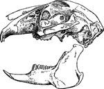
"Terrestril Rodents, with imperfect clavicles, elongated hind limbs, short recurved tail, and long ears. Skull compressed, frontals with large wing-shaped post-orbital processes; molars as in Lagomyidae." —The Encyclopedia Britannica, 1903

Lepus Timidus

"Terrestril Rodents, with imperfect clavicles, elongated hind limbs, short recurved tail, and long ears.…

The membranous labyrinth is lodged within the bony labyrinth and has the same general form; it is, however, considerably smaller and is partly separated from the bony walls by a quantity of fluid, the perilymph. In certain places, it is fixed to the walls of the cavity. The membranous labyrinth contains fluid, the endolymph, and on its walls the ramifications of the acoustic nerve are distributed. Within the osseous vestibule, the membranous labyrinth does not quite preserve the form of the bony cavity, but consists of two membranous sacs, the utricle, and the saccule. The membranous labyrinth is also the location for the receptor cells found in the inner ear.

Membranous Labyrinth

The membranous labyrinth is lodged within the bony labyrinth and has the same general form; it is, however,…

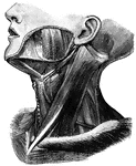
"Principal Muscles on the Left Side of the Neck." — Blaisedell, 1904

Muscles of the neck

"Principal Muscles on the Left Side of the Neck." — Blaisedell, 1904

Diagram to illustrate the action of the ossicles of the middle ear in the conduction of sound to the internal ear.

Ossicles of the Middle Ear

Diagram to illustrate the action of the ossicles of the middle ear in the conduction of sound to the…
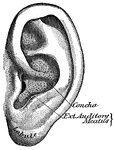
"The outer ear consists of a plate of gristle, shaped somewhat like a shell, known as the pinna, or auricle, and of a tube about an inch long, called the auditory canal." — Blaisedell, 1904

Pinna

"The outer ear consists of a plate of gristle, shaped somewhat like a shell, known as the pinna, or…
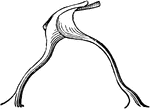
Inner surface of cartilage of pinna.

Inner Surface of Pinna Cartilage

Inner surface of cartilage of pinna.

Outer surface of cartilage of pinna.

Outer Surface of Pinna Cartilage

Outer surface of cartilage of pinna.

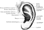
View of outer surface of left pinna.

Outer Surface of Pinna

View of outer surface of left pinna.

The rods of Corti of the ear showing a pair of rods separated from the rest. Labels: i, inner, and e, outer rods.

The Rods of Corti

The rods of Corti of the ear showing a pair of rods separated from the rest. Labels: i, inner, and e,…

The rods of Corti of the ear. Shown hear is a bit of the basilar membrane with several rods on it, showing how they cover in the tunnel of Corti. Labels: i, inner, and e, outer rods;, b, basilar membrane; r, reticular membrane.

The Rods of Corti

The rods of Corti of the ear. Shown hear is a bit of the basilar membrane with several rods on it, showing…

"And she went, and came and gleaned in the field after the reapers: and her hap was to light on the portion of the field belonging unto Boaz, who was of the family of Elimelech. And, behold, Boaz came from Bethlehem, and said unto the reapers, Jehovah be with you. And they answered him, Jehovah bless thee. Then said Boaz unto his servant that was set over the reapers, Whose damsel is this? And the servant that was set over the reapers answered and said, It is the Moabitish damsel that came back with Naomi out of the country of Moab:" Ruth 2:3-6 ASV
<p>Illustration of Ruth gleaning grain in the fields of Boaz. She is on her knees at the feet of Boaz, who walks with a staff and a dog behind him. His servant tells him about Ruth. Four men and women, also gleaning, watch in the background. A small town and some trees are pictured in the background.

Ruth Meets Boaz While Gleaning in the Fields

"And she went, and came and gleaned in the field after the reapers: and her hap was to light on the…
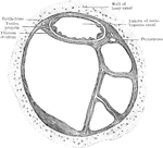
Transverse section of human semicircular canal.

Section of Semicircular Canals

Transverse section of human semicircular canal.

"An ear of wheat with smut, a disease of corn caused by fungi."&mdash;Finley, 1917

Ear of wheat with smut

"An ear of wheat with smut, a disease of corn caused by fungi."—Finley, 1917

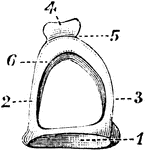
The stapes on stirrup-bone. 1, base; 2 and 3, arch; 4, head of bone, which articulates with orbicular process of the incus; 5, constricted part of the neck; 6, one of the crura.

Stapes on Stirrup-Bone

The stapes on stirrup-bone. 1, base; 2 and 3, arch; 4, head of bone, which articulates with orbicular…
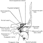
Frontal section of temporal bone, showing the cavities of the outer, middle, and inner ear and the four sides of the petrous.

Frontal Section of Temporal Bone

Frontal section of temporal bone, showing the cavities of the outer, middle, and inner ear and the four…

Section through left temporal bone, showing inner wall of tympanic cavity.

Tympanic Cavity

Section through left temporal bone, showing inner wall of tympanic cavity.

View of the inner wall of the middle ear. Section through the left temporal bone of a child to show the relations of the middle ear and the mastoid antrum to the middle and posterior fossae of the skull.

Inner Wall of Tympanic Cavity

View of the inner wall of the middle ear. Section through the left temporal bone of a child to show…

View of the outer wall of the middle ear. Section through the left temporal bone of a child to show the relations of the middle ear and the mastoid antrum to the middle and posterior fossae of the skull.

Outer Wall of Tympanic Cavity

View of the outer wall of the middle ear. Section through the left temporal bone of a child to show…

Left tympanic membrane (as viewed from the external auditory meatus).

Tympanic Membrane

Left tympanic membrane (as viewed from the external auditory meatus).

Tympanic ossicles of left ear. A, incus as seen from front. B, Malleus, viewed from behind. C, Incus seen from inner aspect. D, Malleus seen from inner aspect. E, Stapes. Labels: 1, body of incus, with articular surface for head of malleus; 2, processus longus; 3, processus lenticularis; 4, articular surface of incus; 5, head; 6, neck; 7, processus brevis; 8, manubrium; 9, body; 10, short process; 11, long process; 12, processus longus; 13, head; 14, facet for incus; 15, manubrium; 16, head; 17, neck; 18, crus anterior; 19, crus posterior; 20, footplate.

Tympanic Ossicles

Tympanic ossicles of left ear. A, incus as seen from front. B, Malleus, viewed from behind. C, Incus…
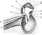
Left membrana tympani and chain of tympanic ossicles.

Tympanic Ossicles

Left membrana tympani and chain of tympanic ossicles.

Interior view of the tympanum, with membrana tympani and bones in natural position. 1, Membrana tympani; 2, Eustachian tube; 3, tensor tympani muscle; 4, lig. mallei super; 6, corda-tympani nerve; a, b, and c, sinuses about ossicula.

Tympanum

Interior view of the tympanum, with membrana tympani and bones in natural position. 1, Membrana tympani;…
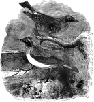
A migratory species of bird, found in Europe, Greenland, and probably North America

Wheatear

A migratory species of bird, found in Europe, Greenland, and probably North America