Two diagrams to illustrate the development of the mesenteries. In the first figure the rotation of the intestinal loop and the continuous primitive mesentery is shown. In the second figure (to the right), which shows a more advances stage, the portions of the primitive mesentery (going to the ascending and descending colons) which disappear through their adhesion to the posterior abdominal wall, are shaded dark, the portions which persist are lightly shaded.

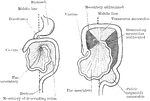
Development of the Mesenteries

Two diagrams to illustrate the development of the mesenteries. In the first figure the rotation of the…

Diagram to illustrate the development of the great omentum. A, shows the beginning of the great omentum and its independence of the transverse mesocolon; in B, the two come in contact; and in C, they have fused along the line of contact. Labels: A, stomach; B, transverse colon; C, small intestine; D, duodenum; E, pancreas; F, great omentum; G, placed in great sac; H, in small sac of peritoneum.

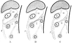
Development of the Great Omentum

Diagram to illustrate the development of the great omentum. A, shows the beginning of the great omentum…

Vertical section of the intestinal mucous membrane of the rabbit. Two villi are represented, in one of which the dilated lacteal alone is shown, in the other the blood vessels and lacteal are both seen injected, the lacteal white, the blood vessels dark; a, the lacteal vessels of the villi; a', horizontal lacteal, which they join; b, capillary blood vessels in one of the villi; c, small artery; d, vein; e, the epithelium covering the villi; g, tubular glands or crypts of Lieberkuhn, some divided down the middle, others cut more irregularly; i, the submucous layer. A, cross-section of three tubular glands more highly magnified.

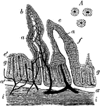
Rabbit's Intestinal Mucous Membrane

Vertical section of the intestinal mucous membrane of the rabbit. Two villi are represented, in one…