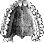
"The head is represented as having been thrown back, and the tongue drawn forward. <em>A</em>, <em>B</em> and <em>C</em>, cainine; <em>D, E</em> bicuspids; <em>F, H, K,</em> molars; <em>M</em> anterior pillar of the fauces; <em>N</em>, tonsil; <em>L</em> uvula; <em>O</em>, upper part of the pharynx; <em>P</em>, tongue drawn forward; <em>R</em> linear ridge, or raphe</em>." &mdash; Blaisedell, 1904

Back view of the adult mouth

"The head is represented as having been thrown back, and the tongue drawn forward. A, B

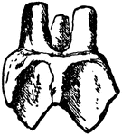
"Side view of second upper molar tooth of Anchitherium (brachyodont form)." &mdash; Encyclopedia Britannica, 1893

Horse Molar

"Side view of second upper molar tooth of Anchitherium (brachyodont form)." — Encyclopedia Britannica,…
"Corresponding tooth of horse (hypsidont form)." &mdash; Encyclopedia Britannica, 1893

Horse Molar

"Corresponding tooth of horse (hypsidont form)." — Encyclopedia Britannica, 1893

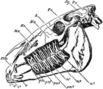
"Side view of skull of horse, with the bone removed so as to expose the whole of the teeth. PMx, premaxilla; Mx, maxilla; Na, nasal bone; Ma, malar bone; L, lacrymal bone; Fr, frontal bone; Sq, squamosal bone; Pa, parietal bone; oc, occipital condyle; pp, paroccipital process; i, the three incisor teeth; c, the canine tooth; pm1, the situation of the rudimentary first premolar, which has been lost in the lower, but is present in the upper jaw; pm2, pm3, and pm4, the three fully-developed premolar teeth; m, the three true molar teeth." &mdash; Encyclopedia Britannica, 1893

Horse Skull

"Side view of skull of horse, with the bone removed so as to expose the whole of the teeth. PMx, premaxilla;…
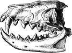
"Dentition of Hyaenodon leptorhynchus. The posterior molar is concealed behind the penultimate tooth." &mdash;The Encyclopedia Britannica, 1903

Hyaenodon Leptorhynchus

"Dentition of Hyaenodon leptorhynchus. The posterior molar is concealed behind the penultimate tooth."…
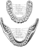
Horizontal section through both the upper and lower jaws to show the roots of the teeth. The sections were carried through the bones a short distance from the edge of their alveolar borders. The upper figure shows the upper teeth, the lower figure the lower teeth. Note the flattened roots of the lower incisors, the two root canals in the anterior root of each lower molar, and the confluence of the three roots of the upper wisdom teeth.

Jaw Showing Roots of Teeth

Horizontal section through both the upper and lower jaws to show the roots of the teeth. The sections…

The molar teeth of a human, horse and dog. The first image to the left in a molar tooth of a horse. Labels: 1, The enamel. 2, The ivory. 3, Canals for blood vessels. The center image is a molar tooth of a human. Label: 1, The enamel. 2, The ivory. 3, The cavity containing blood vessels. 4, 5, Artery and nerve. The image on the right is a molar tooth of a dog. Labels: 1, The enamel. 2, The ivory. 3, Cavity for blood-vessels.

Comparison of the Molar Teeth of a Human, Horse, and Dog

The molar teeth of a human, horse and dog. The first image to the left in a molar tooth of a horse.…

Impaction of the upper third molar in the maxilla. B, Impaction of the lower third molar in the mandible.
Impacted Third Molar

Impaction of the upper third molar in the maxilla. B, Impaction of the lower third molar in the mandible.

Second molar teeth to the left side, labial (A) and lateral (B) aspects.

Second Molar

Second molar teeth to the left side, labial (A) and lateral (B) aspects.

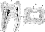
Longitudinal section (A) and transverse section (B) of a human molar tooth. Labels: c, cement; d, dentine; e, enamel; v, pulp cavity

Structure of a Molar

Longitudinal section (A) and transverse section (B) of a human molar tooth. Labels: c, cement; d, dentine;…
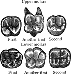
Triturating surfaces of molar teeth of right side. The upper margin of the figures corresponds to the labial surface.

Surface of Molar

Triturating surfaces of molar teeth of right side. The upper margin of the figures corresponds to the…

Vertical section of a molar tooth. Labels: a, enamel of the crown, the line of which indicate the arrangement of its columns; b, dentine; c,cement; d, pulp cavity.

Vertical Section of a Molar

Vertical section of a molar tooth. Labels: a, enamel of the crown, the line of which indicate the arrangement…
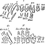
"Milkand permanent dentition of upper (I) and lower (II) jaw of the dog, with the symbols by which the different teeth are commonly designated. The third upper molar (m3) is the only tooh wanting in this animal to complete the typical heterodont mammalian dentition." &mdash;The Encyclopedia Britannica, 1903

Permanent Dentition

"Milkand permanent dentition of upper (I) and lower (II) jaw of the dog, with the symbols by which the…
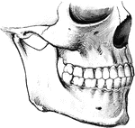
To show the relation of the upper to the lower teeth when the mouth is closed. The manner in which a tooth of one row usually strikes against two teeth of the opposite row, and the resulting interlocking of the teeth, is to be noted.

Teeth

To show the relation of the upper to the lower teeth when the mouth is closed. The manner in which a…

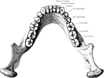
The lower permanent teeth, viewed from above.

Lower Permanent Teeth

The lower permanent teeth, viewed from above.

Permanent teeth, right side.

Permanent Teeth

Permanent teeth, right side.

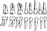
The permanent teeth of the right side, outer or labial aspect. The upper row shows the upper teeth, the lower row the lower teeth. The wide vertical labial ridge is distinct on the upper canine and premolar teeth.

Permanent Teeth

The permanent teeth of the right side, outer or labial aspect. The upper row shows the upper teeth,…

The permanent teeth of the right side, inner of lingual aspect. The upper row shows the upper teeth, the lower row the lower teeth. The cingulum is distinct on the upper incisors and both canines, the lingual cusp on the upper lateral incisor and the upper canine.

Permanent Teeth

The permanent teeth of the right side, inner of lingual aspect. The upper row shows the upper teeth,…
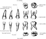
The temporary teeth of the left side. The masticating surfaces of the tow upper molars are shown above. In the second row the upper teeth are viewed from the outer or labial side. In the third row the lower teeth are shown in a similar manner; and below are the masticating surfaces of the two lower molars. In the specimen from which the first upper molar was drawn the two outer or buccal cusps were not distinctly separated, as is often the case.

Temporary Teeth

The temporary teeth of the left side. The masticating surfaces of the tow upper molars are shown above.…
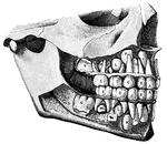
"<em>Temporary Teeth:</em> <em>A</em>, central incisors; <em>B</em>, lateral incisors; <em>C</em>, canines; <em>D</em>, anterior molars; <em>E</em>, posterior molars, <em>F</em>, central incisors; <em>H</em>, lateral incisors; <em>K</em>, canines; <em>L</em>, first bicuspids; <em>M</em>second bicuspids; <em>N</em>, first molars." &mdash; Blaisedell, 1904

Temporary and permanent teeth

"Temporary Teeth: A, central incisors; B, lateral incisors; C, canines;…

The upper permanent teeth.

Upper Permanent Teeth

The upper permanent teeth.

Diagram to illustrate the development of a tooth. I. Shows the downgrowth of the dental lamina D.L. from the surface epithelium E and the beginning of the enamel germ E.G. II. Shows the further growth of the enamel germ and its invagination. III. The enamel germ is more invaginated, and its inner layer of cells becomes columnar. A, the dental lamina, grows thinner, but near its posterior or lingual edge there is an enlargement R.G which is the reserve germ for a permanent tooth. The superficial cells of the dentine papilla P are becoming columnar. IV. The inner columnar cells of the enamel germ ( called enamel cells) A have formed a cap of enamel EN, inside which the superficial cells of the papilla, the odontoblasts O, have formed a layer of dentine D. V. Shows a more advances stage still. The deposit of dentine is extending downwards, and enclosing the papilla to form the future pulp, in which a vessel V is seen. Labels: A, inner layer; B, outer layer; C, remains of intermediate cells; D, dentine; D.L, dental lamina; E, epithelium; E.G, enamel germ; EN, enamel; F, dental furrow; L.D, labio-dental furrow; M, connective tissue cells; O, odontoblasts; P, dentine papilla; R.G, reserve germ; V, blood vessels.

Development of a Tooth

Diagram to illustrate the development of a tooth. I. Shows the downgrowth of the dental lamina D.L.…
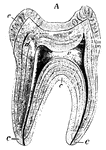
A tooth is generally described as possessing a crown, neck, and root. Side view of a tooth.

Human Tooth

A tooth is generally described as possessing a crown, neck, and root. Side view of a tooth.
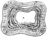
A tooth is generally described as possessing a crown, neck, and root. Top view of a tooth.; 1. Central Nippers, worn to a plain surface.; 2. next pair still sowing a slight remnant of the cavity.; 3. Corner nippers, showing the mark plainly enough.; 4. Tushes, more worn down that in the lower jaw of the six-year-old mouth.

Human Tooth

A tooth is generally described as possessing a crown, neck, and root. Top view of a tooth.; 1. Central…
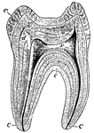
A sectional view of a human molar. The roots, or fangs, are shown covered by a layer of bone called "cement", which is represented by letter c. The center of the tooth is the vascular pulp-cavity v, d is ivory or dentine, and e is the hardest tissue of the body - enamel.

Human Tooth

A sectional view of a human molar. The roots, or fangs, are shown covered by a layer of bone called…