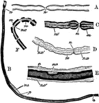
To illustrate the structure of nerve fibers. Labels: A, nerve fiber examined fresh; n, node. B, nerve fiber with axis cylinder shaded, and medulla represented by dark lines; n.c, nucleus; p, granular cell substance near the nucleus. C, more highly magnified: m, medulla; n, node. D, nerve treated with reagents to show the axis cylinder: n.x, surrounded by medulla, m. E, nerve treated with reagents to show n.c, nucleus with fine line over it representing the neurilemma, and outside this fine connective tissue, c: n.c', nucleus lying in the fine connective tissue. F, nerve fiber deprived of its neurilemma showing medulla broken up into fragments, m, surrounding the axis cylinder, n.x.

Nerve Fibers

To illustrate the structure of nerve fibers. Labels: A, nerve fiber examined fresh; n, node. B, nerve…
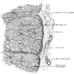
Peripheral part of transverse section of spinal cord, showing nerve fibers subdivided into groups by ingrowth of subpial layer of neuroglia.

Transverse Section of Spinal Cord

Peripheral part of transverse section of spinal cord, showing nerve fibers subdivided into groups by…