Clipart tagged: ‘"optic nerve"’
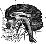
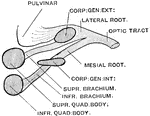
The Cranial Nerves
Diagram of the cranial nerves, 1-4 (Olfactory, Optic, Motor Oculi, and Trochlear).
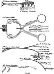
The Eye and its Muscles
The eye and its muscles. Labels: o, the nerve of sight; a, one of the muscles of the eye.
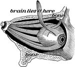
Eye of a Crayfish
A, section through the compound eye of a crayfish. Labels: 1, cornea; 2, crystaline cones; 3, retinulae;…
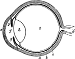
The Eye
The eye. Labels: a, sclerotica; e, cornea; b, choroid; d, optic nerve; f, aqueous humor; g g , iris;…
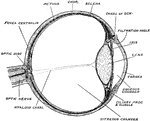
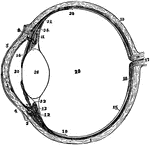
Left Eyeball in Horizontal Section
The left eyeball in horizontal section from before back. Labels: 1, sclerotic; 2, junction of sclerotic…
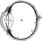
Eyeball
Horizontal section of the eyeball, showing the suspensory ligament of the lens, the aqueous and vitreous…
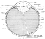
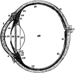
The Eyeball in Horizontal Section
The left eyeball in horizontal section from before back. Labels: 1, sclerotic; 2, junction of sclerotic…
Section of Left Eyeball
The left eyeball in horizontal section from before back. Labels: 1, sclerotic; 2, junction of sclerotic…
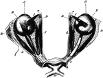
The Eyeballs and Their Muscles
The eyeballs and their muscles as seen when the roof of the orbit has been moved and the fat in the…
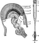
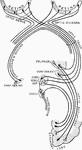
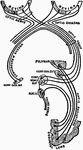
Optic Nerve
The terminal portion of the optic nerve and its entrance into the eyeball, in horizontal section.
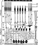
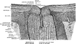
Retinal Structure
Diagram of the structure of the human retina. Labels: I, pigment layer; II, rod and cone layer; R, rods;…