Stomach, liver, pancreas, and duodenum of a dog. Labels: a, liver; b, gall bladder; c, biliary canals; d, cystic duct; e, ductus choledocus; f, pancreas; g, pancreatic ducts.

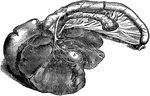
Digestive Organs of a Dog

Stomach, liver, pancreas, and duodenum of a dog. Labels: a, liver; b, gall bladder; c, biliary canals;…
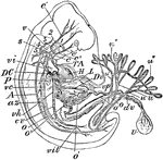
"Early Human Embryo, giving diagrammatically the principal vessels antecedent to the establishment of the regular fetal circulation. H, heart; P, lungs; L, liver; T A, the aortic trunk or cardiac aorta; c, c', c", common, external, and internal carotids; s, subclavian artery; v, vertebral artery; 1, 2, 3, 4, 5, the aortic arches (the persistent left aortic arch hidden); A, subvertebral aorta; o, o', omphalomeseraic artery and vein, to and from U, the umbilical vesicle with its vitelline duct, dv; u, u, the two hypogastric or umbilical arteries, with the ramifications, u", u", in the placenta; u', umbilical vein; vh, hepatic vein; cv, inferior vena cava; vil, iliac veins; az, an azygous vein; vc, a posterior cardinal vein; vi, innominate vein; vp, portal vein; Dv, the ductus venosus; DC, a ductus Cuvieri. The anterior cardinal vein is seen beginning in the head and running down to the ductus Cuvieri, on the under side of the numbers 1, 2, 3, 4, 5." -Whitney, 1911

Human Embryo

"Early Human Embryo, giving diagrammatically the principal vessels antecedent to the establishment of…

The position of the organs of the mouth, thorax, and abdomen. 1, 2, and 3: Salivary glands. 4: The larynx and trachea. 5: The aesophagus. 6: The lungs. 7: The heart. 8: The liver. 9: The stomach. 10: The pancreas. 11: The spleen. 13: The small intestine. 14: The large intestine.

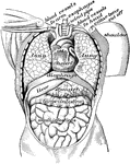
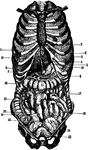
Front View of the Body

The position of the organs of the mouth, thorax, and abdomen. 1, 2, and 3: Salivary glands. 4: The larynx…
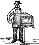
A portable organ played by means of a cylinder set with pins and staples, and turned with a crank.

Hand-organ

A portable organ played by means of a cylinder set with pins and staples, and turned with a crank.

A, the natural position of the internal organs. B, when deformed by tight lacing. The liver and stomach have been forced downward almost as low as the pelvis.

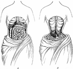
The Natural Position Compared to the Deformed Position of the Internal Organs

A, the natural position of the internal organs. B, when deformed by tight lacing. The liver and stomach…

Organs in the body cavity viewed from the front.

View of Organs from the Front

Organs in the body cavity viewed from the front.

The chief organs of the body from the side. Labels: a, arch of the aorta or main artery of the trunk; c, large intestine; d, diaphragm; e, throat; g, lung; k, kidney; l, spinal cord within the back bone; m, main artery divided to go to h legs; n, pancreas; o, gullet; s, spleen; tr, wind pipe; t, main vein of body; ap, appendix.

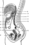
View of Organs from the Side

The chief organs of the body from the side. Labels: a, arch of the aorta or main artery of the trunk;…

The peritoneum is a large serous membrane, which forms in the male a closed sac, the parietal layer of which lines the abdominal walls, its visceral layer being reflected more or less completely over all the abdominal and pelvic viscera. It's free surface is covered with endothelium, and is smooth, moist, and shining. Its attached surface is connected to the viscera and the parietes of the abdomen by the sub-peritoneal tela or areolar tissue. In the female it is not a closed sac, the free extremities of the Fallopian tubes opening directly into the cavity. The peritoneum is divided by a constricted portion of its tissue, at the foramen of Winslow into 2 sacs, the Greater Sac and the Lesser Sac.

The Peritoneum

The peritoneum is a large serous membrane, which forms in the male a closed sac, the parietal layer…

A side view of the two great cavities of the body and their organs. 1: The mouth. 2: The thorax. 3: The abdomen. 4: The brain. 5: The spinal cord. 6: A salivary gland. 7: The aesophagus. 8: The trachea and lungs. 9: The heart. 10: The liver. 11: The stomach. 12: The colon. 13: The small intestine. 14: The lacteals. 15: The thoracic duct. 16: The diaphragm. 17, 18, and 20: The walls of the lower cavity, composed of bones, muscles and skin. 19 and 20: The walls of the upper cavity.

Side View of the Body

A side view of the two great cavities of the body and their organs. 1: The mouth. 2: The thorax. 3:…

Diagram showing the position of the thoracic and abdominal organs. labels: 1, lower border of right lung; 2, the same of the left lung; 3, liver, right lobe; 4, liver, left lobe; 5, suspensory ligament of the liver; 6, fundus of gall-bladder; 7, cardia of stomach; 8, fundus of stomach; 9, lower border of stomach; 10, position of pylorus; 11, caecum; 12, vermiform appendix; 13, ascending colon; 14, right flexure of colon; 15, transverse colon, concealed by, 19, convolutions of the small intestine; 20, termination of ileum, ascending from right to left; 21, bladder, distended, partly covered by peritoneum; 22, the part of the bladder which is not covered by peritoneum.

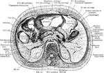
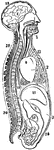
Section Across the Forearm

Diagram showing the position of the thoracic and abdominal organs. labels: 1, lower border of right…
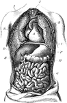
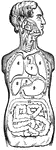
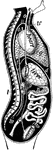
The human torso. Labels: A, the heart; B, the lungs drawn aside to show the internal organs; C, the diaphragm; D, the liver; E, the gall cyst; F, the stomach; G, the small intestines; H, the transverse colon.

Torso

The human torso. Labels: A, the heart; B, the lungs drawn aside to show the internal organs; C, the…

Frozen section across body at level of twelfth thoracic vertebra.

Section Across Trunk

Frozen section across body at level of twelfth thoracic vertebra.