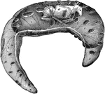
Fetus of a cow, with its membrane. Labels: a, placenta; b, chorion with the allantois adherent to its inner surface; c, amnion seen through outer covering; d, fetus seen through its covering.

Cow Fetus

Fetus of a cow, with its membrane. Labels: a, placenta; b, chorion with the allantois adherent to its…

Fetus of a dog, with its membrane. Labels: a, placenta; b, chorion with the allantois adherent to its inner surface; c, amnion seen through outer covering; d, fetus seen through its covering.

Dog Fetus

Fetus of a dog, with its membrane. Labels: a, placenta; b, chorion with the allantois adherent to its…
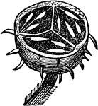
The inside of a gooseberry, showing the reproductive seeds that are immersed in the placenta.

Gooseberry

The inside of a gooseberry, showing the reproductive seeds that are immersed in the placenta.
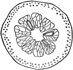
"The Hesperidium is a fleshy fruit, in which the epicarp and mesocarp form a thick rind, and the endocarp projects inward, forming triangular divisions, in which pulpy cells are developed, which surround the seeds."—Darby, 1855

Hesperidium

"The Hesperidium is a fleshy fruit, in which the epicarp and mesocarp form a thick rind, and the endocarp…

"The Pepo is an inferior fruit, with a thick and fleshy rind, with two or more fleshy parietal placentas, projecting more or less inward."—Darby, 1855

A Section of the Pepo

"The Pepo is an inferior fruit, with a thick and fleshy rind, with two or more fleshy parietal placentas,…
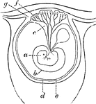
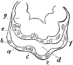
Diagram of an early stage of the formation of the human placenta. Labels: a, embryo; b, amnion; c, placental vessels; d, decidua reflexa; e, allantois; f, placental villi; g, mucous membrane.

Early Formation of the Placenta

Diagram of an early stage of the formation of the human placenta. Labels: a, embryo; b, amnion; c, placental…

Section of human placenta at end of pregnancy. The fetal blood vessels have been injected; the maternal blood spaces appear as clear areas surrounding the sections of the fetal villi.

Section of Placenta

Section of human placenta at end of pregnancy. The fetal blood vessels have been injected; the maternal…
Extremities of a placental villus. Labels: a, lining membrane of the vascular system of the mother; b, cells immediately lining a; d, space between the maternal and fetal portion of the villus; e, internal membrane of the chorion; f, internal cells of the villus, or cells of the chorion; g, loop of umbilical vessels.

Placental Villus

Extremities of a placental villus. Labels: a, lining membrane of the vascular system of the mother;…

"The Silicua is a variety of the capsule, composed of two carpels opening from the base upward, and having a framework called the replum, which forms a spurious dissepiment."—Darby, 1855

Silicula

"The Silicua is a variety of the capsule, composed of two carpels opening from the base upward, and…
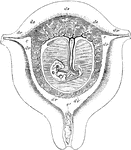
Diagrammatic view of a vertical transverse section of the uterus at the seventh week of pregnancy. Labels: c, c', cavity of uterus, which becomes the cavity of the decidua, opening at c, the cornua, into the Fallopian tubes, and at c' into the cavity of the cervix, which is closed by a plug of mucus; dv, decidua vera; dr, decidua reflexa, with the sparser villi imbedded in its substance; ds, decidua serotina, involving the more developed chorionic villi of the commencing placenta. The fetus is seen lying in the amniotic sac; passing up from the umbilicus is seen the umbilical pedicle of the yolk sac, which lies in the cavity between the amnion and chorion.

Uterus at Seventh Week of Pregnancy

Diagrammatic view of a vertical transverse section of the uterus at the seventh week of pregnancy. Labels:…