Lateral view of the spinal column. Labels: 1, atlas; 2, dentata 3, seventh cervical vertebra; 4, twelfth dorsal vertebra; 5, fifth lumbar vertebra; 6, first piece of sacrum; 7, last piece of sacrum; 8, coccyx; 9, a spinous process; 10, 10, intervertebral foramina.

Spinal Column

Lateral view of the spinal column. Labels: 1, atlas; 2, dentata 3, seventh cervical vertebra; 4, twelfth…

The spinal column.

Spinal Column

The spinal column.

Lateral view of the vertebral column.

Spinal Column

Lateral view of the vertebral column.

A section of the brain and spinal column. Labels: 1, The cerebrum (large brain). 2, The cerebellum (small brain). 3, The Medulla Oblongata. 4, The spinal cord in its canal.

The Spinal Column and Brain

A section of the brain and spinal column. Labels: 1, The cerebrum (large brain). 2, The cerebellum (small…

Side view of the spinal column. Labels: C 1-7, cervical; D 1-12, dorsal; L 1-15 lumbar; S 1, sacrum; Co 1-4, coccygeal.

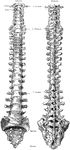
Side View of the Spinal Column

Side view of the spinal column. Labels: C 1-7, cervical; D 1-12, dorsal; L 1-15 lumbar; S 1, sacrum;…
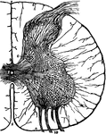
Diagrammatic view from before of the spinal cord and medulla oblongata, including the roots of the spinal and some of the cranial nerves, and on one side of the gangliated chain of the sympathetic. The spinal nerves are enumerated in order on the right side of the figure. Labels: Br, brachial plexus; Cr, anterior crural, O, obturator, and Ss, great sciatic nerves, coming off from lumbo-sacral plexus; x, x, filum terminale; a,b,c, superior, middle, and inferior cervical ganglia of the sympathetic, the last united with the first thoracic, d; d', the eleventh thoracic ganglion; l, the twelfth thoracic (or first lumbar); below ss, the chain of sacral ganglia.

Spinal Cord

Diagrammatic view from before of the spinal cord and medulla oblongata, including the roots of the spinal…

Diagrams of spinal cord and nerve roots. Labels: A, a small portion of the cord seen from the ventral side; B, the same seen laterally; C, a cross-section of the cord; D, the two roots of a spinal nerve; 1, anterior (ventral) fissure; 2, posterior (dorsal) fissure; 3, surface groove along the line of attachment of the anterior nerve roots; 4, line of origin of the posterior roots; 5, anterior root filaments of a spinal nerve; 6, posterior root filaments; 6', ganglion of the posterior root; 7, 7', the first two divisions of the nerve trunk after its formation by the union of the two roots.

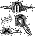
Spinal Cord and Nerve Roots

Diagrams of spinal cord and nerve roots. Labels: A, a small portion of the cord seen from the ventral…

A thin transverse section of half the spinal cord magnified about 10 diameters. Labels: 1, anterior fissure; 2, posterior fissure; 3, central canal ;8, pia mater enveloping the cord; 6,7, bands of pia mater penetrating the cord and supporting its nerve elements; 9, a posterior root; 10, bundles of an anterior root; a,b,c,d,e, groups of nerve cells in the gray matter.

Spinal Cord Section

A thin transverse section of half the spinal cord magnified about 10 diameters. Labels: 1, anterior…

Anterior and posterior view of adult spine.

Anterior and Posterior Views of Spine

Anterior and posterior view of adult spine.

Lateral view of adult spine.

Lateral View of Spine

Lateral view of adult spine.

Lateral and posterior views of the vertebral column.

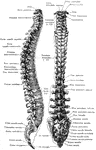
Lateral and Posterior View of the Vertebral Column

Lateral and posterior views of the vertebral column.