Clipart tagged: ‘Squamosal’
Cat Ear
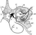
"Diagram showing the ear and related parts in a young cat. P., Pinna; Sq., squamosal: E.A.M., external…
Ripe Chick's Skull Profile
"Ripe chick's skull, longitudinal section, vied inside, x 3 diameters; after Parker. px, premaxillary;…
Ripe Chick's Skull
"Ripe chick's skull, longitudinal section, vied inside, x 3 diameters; after parker. In the mandible…
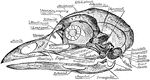
Skull of a Common Fowl
"Fig. 62 Skull of common fowl, enlarged. from nature by Dr. R.W. Shufeldt, U.S.A. The names of bones…
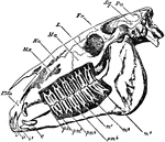
Horse Skull
"Side view of skull of horse, with the bone removed so as to expose the whole of the teeth. PMx, premaxilla;…
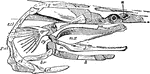
Polyodon
"Skull of Polyodon. n, nasal cavity; sq, squamosal; mh, hyomandibular; sy, symplectic; pa, palate-pterygold;…