Clipart tagged: ‘tapetum’
Angiosperm
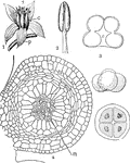
This illustration shows the flower and sporophylls of Angiosperms: 1, flower of Sedum with leaf-like…
Microspore Cells
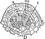
The sporogenous cells (s), tapetum (t), two parietal layers (oo) in the stages of "formation of anthers…
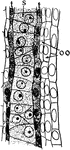
Microspore Cells
The sporogenous cells (s), tapetum (t), two parietal layers (oo) in the stages of "formation of anthers…