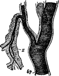
Part of a tracheal stem and branches of an insect. Labels: Z, cellular outer wall; SP, cuticular inner wall with spiral fiber.

Tracheal Stem and Branches

Part of a tracheal stem and branches of an insect. Labels: Z, cellular outer wall; SP, cuticular inner…

Tracheal system of a fly larva. Labels: Tr, longitudinal stem of right side; St', St", anterior and posterior stigmata.

Tracheal System of Fly Larva

Tracheal system of a fly larva. Labels: Tr, longitudinal stem of right side; St', St", anterior and…
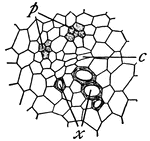
Young vascular bundle: p, primary phloem; x, primary xylem; c, first divisions of cambium cells.

Vascular Bundle

Young vascular bundle: p, primary phloem; x, primary xylem; c, first divisions of cambium cells.
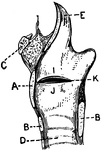
This illustration shows the vocal cords as seen from above during phonation (A. Thyroid Cartilage; B. Cricoid Cartilage; C. hyoid bone; D. Rings of trachea; E. Epiglottis; F. Arytenoid Cartilage; G. Thyro-arytenoid muscle; H. Adam's Apple; I. False Vocal Cords; J. True Vocal Cords; K. Ventricle; L. Rima Glottis).

Vocal Cords Seen from above During Phonation

This illustration shows the vocal cords as seen from above during phonation (A. Thyroid Cartilage; B.…
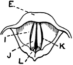
This illustration shows the vocal cords, seen from above during quiet breathing (A. Thyroid Cartilage; B. Cricoid Cartilage; C. hyoid bone; D. Rings of trachea; E. Epiglottis; F. Arytenoid Cartilage; G. Thyro-arytenoid muscle; H. Adam's Apple; I. False Vocal Cords; J. True Vocal Cords; K. Ventricle; L. Rima Glottis).

Vocal Cords Seen from above During Quiet Breathing

This illustration shows the vocal cords, seen from above during quiet breathing (A. Thyroid Cartilage;…
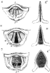
Three laryngoscopic view of the superior aperture of the larynx and surrounding parts. Labels: A, the glottis during the emission of a high note in singing; B, in easy and quite inhalation of air; C, in the state of widest possible dilation, as in inhaling a very deep breath. The diagrams A', B', and C', show in horizontal sections of the glottis in position of the vocal ligaments and arytenoid cartilages in the three several states represented in the other figures. In all the figures, so far as marked, the letters indicate the parts as follows, viz. : l, the base of the tongue; e, the upper free part of the epiglottis; e', the tubercle or cushion of the epiglottis; p h, part of the anterior all of the pharynx behind the larynx; in the margin of the aryteno-epiglottisdean fold w, the swelling of the membrane caused by the cartilages of Wrisberg; s. that of the cartilages of Santorini: a, the tip or summit of the arytenoid cartilages; c v, the true vocal cords r lips of the rima glottidis; c v s, the superior or false vocal cords; between them the ventricle of the larynx; in C, tr, is placed on the anterior wall of the receding trachea, and b indicated the commencement of the two bronchi beyond the bifurcation which may be brought into view in this state of extreme dilation.

Movement of the Vocal Cords

Three laryngoscopic view of the superior aperture of the larynx and surrounding parts. Labels: A, the…
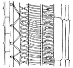
Longitudinal section (diagrammatic) of a young xylem strand; c, cambium; y, young trachea,; p, pitted trachea; s, spiral trachea; a, annular trachea; m, pith.

Xylem

Longitudinal section (diagrammatic) of a young xylem strand; c, cambium; y, young trachea,; p, pitted…