Clipart tagged: ‘Uterus’
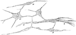
Chorion Villi
Very soon after the entrance of the ovum into the uterus, in the human subject, the outer surface of…
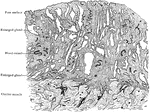
Mucous Membrane of Uterus in Fourth Month of Pregnancy
Section of mucous membrane lining body of uterus (decidua vera); fourth month of pregnancy.
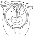
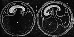
Early Formation of the Placenta
Diagram of an early stage of the formation of the human placenta. Labels: a, embryo; b, amnion; c, placental…
Tissue Growth of the Uterus
In the embryo the place of fibrous tissues is at first occupied by a mass of roundish cells, derived…
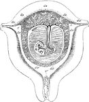
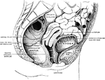
Uterus at Seventh Week of Pregnancy
Diagrammatic view of a vertical transverse section of the uterus at the seventh week of pregnancy. Labels:…
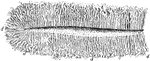
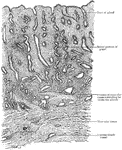
Lining Membrane of the Uterus
Section of the lining membrane of a human uterus at the period of commencing pregnancy showing the arrangement…