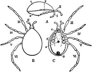
Holothyrus Nitidissimus

“Holothyrus nitidissimus, one of the Acari; ater Thorell. A, Lateral view with appendages III to VI removed, I, plate covering the whole dorsal area, representing the fused tergal sclerites of the prosoma and opisthosoma; 2, similarly-formed ventral plate; 3, tracheal stigma. B, dorsal view of the same animal; II to VI, 2nd to 6th pairs of appendages. The 1st pair of appendages both in this and in C are retracted. C, Ventral view of the same; II to VI as in B; a, genital orifice; b, anus; c, united basal segments of the second pair of appendages; d, basal segment of the 6th prosomatic appendage of the right side. The rest of the appendage, as also of app. III, IV and V, has been cut away.” — The Encyclopedia Britannica, 1910
Galleries
Spiders, Mites and ScorpionsSource
The Encyclopedia Britannica, Eleventh Edition (New York: The Encyclopedia Britannica Company, 1910)II:310
Downloads
2400×1882, 384.8 KiB
1024×802, 57.2 KiB
640×501, 32.3 KiB
320×250, 13.1 KiB