Peyer's Patch
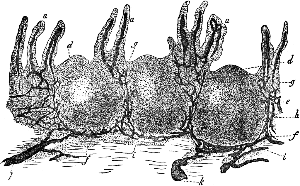
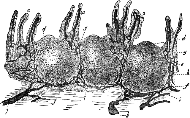
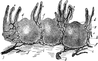
Vertical section of a portion of a Peyer’s Patch, with lacteal vessels injected. Labels: a, villi, with their lacteals colored black; d, surface of rounded follicle, or solitary gland; e, central part; f,g,h,i, and k, lymph-channels, or lacteal vessels, colored black. “Closely connected with the lymphatic vessels in the intestines are small, rounded bodies of the size of a small pin’s head, called solitary glands or follicles. These bodies consist of a rounded mass of fine lymphoid tissue, the meshes of which are crowded with leucocytes. A Peyer’s patch, or agminated gland, as it is often called, is simply a collection of these follicles."— Kimber, 1907
Galleries
Human Lymphatic SystemSource
Kimber, Diana C. Anatomy and Physiology for Nurses (New York, NY: The Macmillan Company, 1907)
Downloads
2400×1488, 1.9 MiB
1024×634, 245.9 KiB
640×396, 108.6 KiB
320×198, 30.0 KiB