This human anatomy ClipArt gallery offers 46 illustrations of the lymphatic system. The lymphatic system is a network for vessels that carry a clear liquid known as lymph that is used in immune functions, as well as transportation of fluids and fatty acids. It also includes lymphoid tissue through which the lymph travels, as well as structures dedicated to the circulation and production of lymphocytes (e.g., spleen, thymus, bone marrow).

"Among the cells of the body there is, besides the blood capillaries, a system of fine, thin-walled lymphatic capillaries." — Ritchie, 1918

Lymphatic Vessels in the Fingers

"Among the cells of the body there is, besides the blood capillaries, a system of fine, thin-walled…
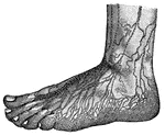
"In nearly every tissue of the body there is a marvelous network of vessels, precisely like the lacteals, known as the <em>lymphatics</em>. They seem to start out of the part in which they are found, like the rootlets of a plant in the soil. The tiny roots join together and make larger roots. They carry a fluid called <em>lymph</em>, very much like blood without the red corpuscles. It is to be remembered that the lacteals are really the lymphatics which begin in the villi of the small intestine." — Blaisedell, 1904

Superficial Lymphatics of the foot

"In nearly every tissue of the body there is a marvelous network of vessels, precisely like the lacteals,…

Lymphatic vessels of the dorsal surface of the hand.

Lymphatic Vessels of the Hand

Lymphatic vessels of the dorsal surface of the hand.

Superficial lymphatics of the digits and of the dorsal aspect of the hand.

Lymphatics of the Hand

Superficial lymphatics of the digits and of the dorsal aspect of the hand.

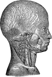
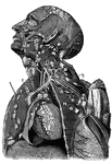
Lymphatics and lymphatic glands of the head and neck.

Lymphatics of the Head and Neck

Lymphatics and lymphatic glands of the head and neck.

End of Lacteal, showing wall and valves, greatly enlarged.

Lacteal

End of Lacteal, showing wall and valves, greatly enlarged.

The lacteals and lymphatics.

The Lacteals and Lymphatics

The lacteals and lymphatics.

A side view of the lacteal and thoracic duct. Labels: 1, Small intestine. 2, Lacteals. 3, Thoracic duct. 4, Stomach. 5, Colon. 6, Pancreas. 7, Liver. 8, Diaphragm. 9 Heart. 10, Lungs. 11, Large vein into which the thoracic duct opens. 12, Spinal column.

A Side View of the Lacteals and Thoracic Duct

A side view of the lacteal and thoracic duct. Labels: 1, Small intestine. 2, Lacteals. 3, Thoracic duct.…
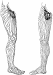
The lymphatics in the leg, with glands at the hip.

Lymphatics in the Leg

The lymphatics in the leg, with glands at the hip.

Superficial lymphatic vessels and nodes of the right lowers extremity and groin.

Superficial Lymphatics and Vessels and Nodes of the Legs

Superficial lymphatic vessels and nodes of the right lowers extremity and groin.

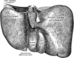
Lymphatics of the inferior surface of the liver.

Lymphatics of the Liver

Lymphatics of the inferior surface of the liver.

The regions whose lymph flows into the right lymphatic duct are suggested by the lighter area; those which are tributary to the thoracic duct by the darker area.

Regions of Lymph Flow

The regions whose lymph flows into the right lymphatic duct are suggested by the lighter area; those…
A lymph node with its afferent and efferent vessels.

Lymph Node

A lymph node with its afferent and efferent vessels.

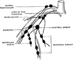
An axillary node is a lymph node of the upper extremity. Shown is the scheme of the axillary nodes. The dotted line indicates the position of the clavicle.

Lymph Node of Upper Extremity

An axillary node is a lymph node of the upper extremity. Shown is the scheme of the axillary nodes.…

A lymph node with its afferent and efferent vessels.

Vessels of a Lymph Node

A lymph node with its afferent and efferent vessels.

"The lymph vessels of the body. rc, the thoracic duct; lac, the lacteals taking the lymph and fatty part of food from the intestines." —Davison, 1910

Lymph Vessels

"The lymph vessels of the body. rc, the thoracic duct; lac, the lacteals taking the lymph and fatty…

The lymph vessels pass through bodies named lymph nodes or glands.

Lymph Vessels and Lymph Nodes

The lymph vessels pass through bodies named lymph nodes or glands.

A lymphatic gland, with several vessels passing through it.

A Lymphatic Gland

A lymphatic gland, with several vessels passing through it.

"Consisting of a very fine network of fibrils, around which are cells of various sizes." — Blaisedell, 1904

Connective Tissue from a Lymphatic Gland

"Consisting of a very fine network of fibrils, around which are cells of various sizes." — Blaisedell,…

Diagrammatic section of lymphatic gland. Labels: a.l., afferent lymphatic; e.l., efferent lymphatic; c, capsule, or envelope; tr, trabeculae; l.s., lymph-sinus; l.h., pulpy substance of gland.

Diagrammatic Section of a Lymphatic Gland (Lymph Node)

Diagrammatic section of lymphatic gland. Labels: a.l., afferent lymphatic; e.l., efferent lymphatic;…
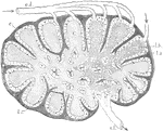
Diagrammatic section of lymphatic gland. a.l., afferent; e.l., efferent lymphatics: C, cortical substance; l.h., reticulating cords of medullary substance; l.s., lymph-sinus; c, fibrous coat sending in trabeculae; t.r., into the substance of the gland.

Diagrammatic Section of Lymphatic Gland

Diagrammatic section of lymphatic gland. a.l., afferent; e.l., efferent lymphatics: C, cortical substance;…
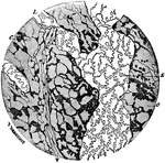
A small portion of a lymphatic plexus, magnified 110 diameters. Labels: L, lymphatic vessel with characteristic endothelium; C, cell spaces of the connective tissue abutting here and there against the lymphatics.

Lymphatic Plexus

A small portion of a lymphatic plexus, magnified 110 diameters. Labels: L, lymphatic vessel with characteristic…

The valves of a lymphatic trunk.

The Valves of a Lymphatic Trunk

The valves of a lymphatic trunk.

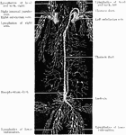
View of the great lymphatic trunks. Labels: 1, 2 Thoracic duct. 4, The right lymphatic duct. 5, Lymphatics of the thigh. 6, Iliac lymphatics. 7, Lumbar lymphatics. 8, Intercostal lymphatics. a, Descending cava. b, Left innominate vein. c, Right innominate vein. d, Aorta. e, Ascending cava.

View of the Great Lymphatic Trunks

View of the great lymphatic trunks. Labels: 1, 2 Thoracic duct. 4, The right lymphatic duct. 5, Lymphatics…
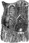
Valves of lymphatics.

Lymphatic Valves

Valves of lymphatics.

A single lymphatic vessel, much magnified.

A Single Lymphatic Vessel

A single lymphatic vessel, much magnified.

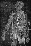
Diagram of the principle groups of the Lymphatic vessels.

Lymphatic Vessels

Diagram of the principle groups of the Lymphatic vessels.

Femoral iliac and aortic lymphatic vessels and glands. Labels: 1, saphena magna vein; 2, external iliac artery and vein; 3, primitive iliac artery and vein; 4, aorta; 9, superior set of inguinal lymphatic glands; 10, chain of lymphatics; 11, lymphatics which accompany the circumflex iliac vessels; 12, lumbar an aortic lymphatics; 13, origin of thoracic duct; 14, thoracic duct at its commencement.

Lymphatic Vessels and Glands

Femoral iliac and aortic lymphatic vessels and glands. Labels: 1, saphena magna vein; 2, external iliac…

The lymphatic vessels and glands. Labels: 1, 2, 3, 4, 5, 6, Lymphatic vessels and glands of the lower limb. 7, Lymphatic glands. 8, The receptacle of the thoracic duct. 9, The lymphatics of the kidney. 10, The stomach. 11, Of the liver. 12, Of the lungs. 13, 14, 15, The lymphatics and glands of the arm. 16, 17, 18, Of the face and neck. 19, 20, Large veins. 21, The thoracic duct.

The Lymphatic Vessels and Glands

The lymphatic vessels and glands. Labels: 1, 2, 3, 4, 5, 6, Lymphatic vessels and glands of the lower…
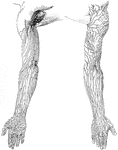
Superficial lymphatic vessels and nodes of the upper extremity.

Lymphatic Vessels and Nodes of the Arms

Superficial lymphatic vessels and nodes of the upper extremity.
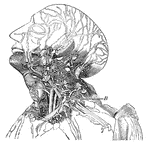
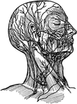
This diagram shows the lumphatics of the head and neck. It shows the glands, and B, the thoracic duct as it empties into the left innominate vein at the junction of the left jugular and subclavian veins.

Lymphatics

This diagram shows the lumphatics of the head and neck. It shows the glands, and B, the thoracic duct…

Superficial lymphatics of the right arm. 1: Vein; 2: Lymphatic tubes; 3: Lymphatic glands.

Lymphatics

Superficial lymphatics of the right arm. 1: Vein; 2: Lymphatic tubes; 3: Lymphatic glands.
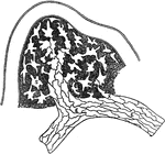
Origin of lymphatics. Labels: S, lymph spaces communicating with lympathic vessel; A, origin of lymphatic by union of lymp spaces; E, E, endothelial cells forming wall of lymph vessel.

Lymphatics

Origin of lymphatics. Labels: S, lymph spaces communicating with lympathic vessel; A, origin of lymphatic…

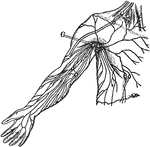
Superficial lymphatics and glands. Labels: G. Axillary glands.

Lymphatics

Superficial lymphatics and glands. Labels: G. Axillary glands.

The lymphatic vessels. The thoracic duct occupies the middle of the figure. it lies upon the spinal column, at the sides of which are seen portions of the ribs (1). Labels: a, the receptacle of the chyle; b, the trunk of the thoracic duct, opening at c, into the junction of the left jugular (f) and subclavian (g) veins as they unite into the left innominate vein, which has been cut across to show the thoracic duct running behind it; d, lymphatic glands placed in the lumbar regions; h, the superior vena cava formed by the junction of the right and left innominate veins.

Lymphatics

The lymphatic vessels. The thoracic duct occupies the middle of the figure. it lies upon the spinal…

Lymphatics and lymphatic glands (lymph nodes) of axilla and arm.

Lymphatics and Lympatic Glands (Lymph Nodes) of Axilla and Arm

Lymphatics and lymphatic glands (lymph nodes) of axilla and arm.

Superficial lymphatics of right groin and upper part of thigh. Labels: 1, Upper inguinal glands. 2, 2'. Lower or inguinal or femoral glands. 3, 3'. Plexus of lymphatics in the course of the long saphenous vein.

Lymphatics of Groin and Thigh

Superficial lymphatics of right groin and upper part of thigh. Labels: 1, Upper inguinal glands. 2,…

Lymphatic vessels near the surface of the skin.

Superficial Lympatics

Lymphatic vessels near the surface of the skin.

Vertical section of a portion of a Peyer's Patch, with lacteal vessels injected. Labels: a, villi, with their lacteals colored black; d, surface of rounded follicle, or solitary gland; e, central part; f,g,h,i, and k, lymph-channels, or lacteal vessels, colored black. "Closely connected with the lymphatic vessels in the intestines are small, rounded bodies of the size of a small pin's head, called solitary glands or follicles. These bodies consist of a rounded mass of fine lymphoid tissue, the meshes of which are crowded with leucocytes. A Peyer's patch, or agminated gland, as it is often called, is simply a collection of these follicles."&mdash; Kimber, 1907

Peyer's Patch

Vertical section of a portion of a Peyer's Patch, with lacteal vessels injected. Labels: a, villi, with…
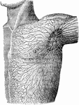
Lymphatics and lymphatic glands on the left side of the body and shoulder.

Lymphatics of the Shoulder

Lymphatics and lymphatic glands on the left side of the body and shoulder.
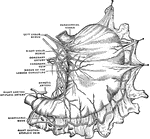
Lymphatics of the small intestine.

Lymphatics of the Small Intestine

Lymphatics of the small intestine.

The spleenvisceral aspect.

Spleen

The spleenvisceral aspect.

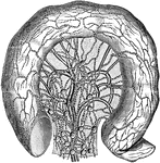
General view of the subperitoneal lymphatic plexus of the stomach prepared by the method of Gerota.

Lymphatic Plexus of the Stomach

General view of the subperitoneal lymphatic plexus of the stomach prepared by the method of Gerota.

"Human Thoracic Duct and Azygous Veins. a, receptacle of the chyle; b, trunk of the thoracic duct, opening at c into root of left innominate vein at junction of f, left jugular, and g, left subclavian vein; e, right innominate vein; d, d, d, several thoracic and lumbar lymphatic glands; h, h, a short portion of the esophagus. Two azygous veins run parallel with and on each side of the duct, until the left crosses behind the duct to join the right. The structures represented rest nearly upon the back-bone." -Whitney, 1911

Thoracic Duct

"Human Thoracic Duct and Azygous Veins. a, receptacle of the chyle; b, trunk of the thoracic duct, opening…
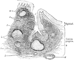
A tonsil consists of an elevation of the mucous membrane presenting 12 to 15 orifices which lead into crypts or recesses, in the walls of which are placed nodules of adenoid or lymphoid tissue. Shown is a vertical section through a crypt of the human tonsil. Labels: 1, entrance to the crypt; 2 and 3, the framework or adenoid tissue; 4, the enclosing fibrous tissue; a and b, lymphatic follicles; 5 and 6, blood vessels.

Crypt of Tonsil

A tonsil consists of an elevation of the mucous membrane presenting 12 to 15 orifices which lead into…

Lymphatic vessels are used to transfer lymph throughout the body.

Lymphatic Vessels

Lymphatic vessels are used to transfer lymph throughout the body.