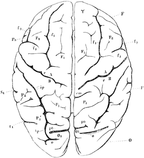
View of Brain from Above

View of brain from above. F, Frontal lobe; P, Parietal lobe; O, Occipital lobe; T, Temporal lobe; S, fissure of Sylvius; S’, horizontal; S", ascending ramus of the same; c, sulcus centralis (fissure of Rolando); A, ascending frontal convolutions; fr, superior, f2, inferior frontal sulcus; f3, precentral sulcus; P1, superior parietal lobule; P2, inferior parietal lobule consisting of P2, supramarginal gyrus, and P2’, angular gyrus; ip, interparietal sulcus; cm, termination of callosomarginal fissure; O1, first O2, second, 03, third occipitals inferior; T1, first T2, second, T3, third temporal convolutions; tr, first , t2, second temporal fissures. Sr, end of horizontal ramus of fissure of Sylvius.
Keywords
brainGalleries
Human Central Nervous SystemSource
Baker, W. Morrant & Harris, Vincent Dormer Kirkes' Hand-book of Physiology, 13th ed. (Philadelphia: P. Blakiston's Son & Co., 1892) 637
Downloads
2400×2628, 666.4 KiB
935×1024, 82.4 KiB
584×640, 41.5 KiB
292×320, 15.7 KiB