This human anatomy ClipArt gallery offers 265 illustrations of the central nervous system, including external and dissected views of the brain and spinal cord.
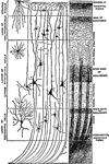
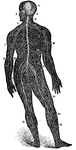
Reflex Action
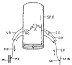
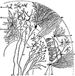
Illustration of the reflex action of an animal. SE is the sensory nerve-ending. A stimulus passes through…
Myelinic Axons
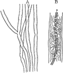
A, Myelinic axons in fresh state, showing few nodes. B, Portion of a myelinic axon treated with boiling…
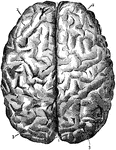
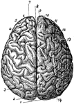
Brain
"Profile and vertex views of cerebrum. Dr, the frontal lobe; Par, parietal; Oc, occipital; Ts, temporo-sphenoidal…
Brain
"Diagram illustrating the general relationships of the parts of the brain. A, fore-brain; b, midbrain;…
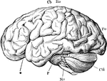
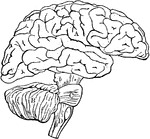
Brain
"The brain from the left side. Cb, the cerebral hemispheres forming the main bulkl of the fore-brain;…
Brain
"Diagram of the left half of a vertical median section of the brain. H, H, convoluted inner surface…
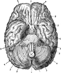
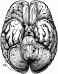
Brain
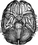
The brain seen from below. 1: Great fissure; 2: Anterior lobes of cerebrum; 3: Posterior lobes of cerebrum;…
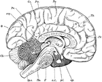
Brain
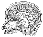
Mesial section through the Corpus Callosum, the Mesencephalon, the Pons, Medulla and Cerebellum. Showing…
Brain
Transverse section through the human mesencephalon at the level of the superior quadrigeminal body


Brain
Orbital surface of the left frontal lobe and the island of Reil; the tip of the temporo-sphenoidal lobe…

Brain
Horizontal section through the Right Cerebral Hemisphere at the Level of the Widest Part of the Lenticular…
Brain
Diagram illustrating the general relationships of the parts of the brain. Labels: A, fore-brain; b,…
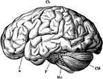
Brain
The brain from the left side. Labels: Cb, the cerebral hemispheres forming the main bulk of the fore-brain;…
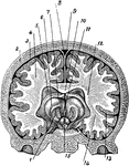
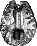
Brain
A cross section of the brain from left to right. Labels: 1, thalamus; 2, skull; 3, cerebral membrane;…
Brain and Cranial Nerves
The brain and the cranial nerves seen partly in section and partly in side view. Labels: C, convolutions…
Brain and Spinal Cord
Anterior view of the brain and spinal marrow. Labels: 1, 1, hemispheres of the cerebrum; 2, great middle…
Brain and Spinal Cord
The brain and spinal cord. Labels: 1, 1, hemispheres of cerebrum; 2, great middle fissure; 3, cerebellum;…
A Back View of the Brain and Spinal Cord
A back view of the brain and spinal cord. Labels: 1, The cerebrum. 2, The cerebellum. 3, The spinal…
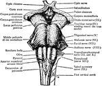
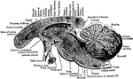
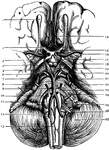
The Brain and the Cranial Nerves
The brain and the origin of the twelve pairs of cranial nerves. Labels: F, E, the cerebrum; D, the cerebellum,…
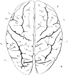
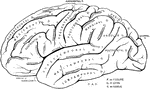
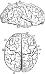
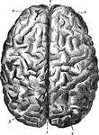
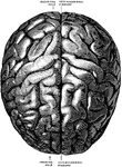
View of Brain from Above
View of brain from above. F, Frontal lobe; P, Parietal lobe; O, Occipital lobe; T, Temporal lobe; S,…
Brain Hemispheres and Spinal Cord
1. Hemispheres of the brain proper, or cerebrum. 2. Hemispheres of the smaller brain, or cerebellum.…
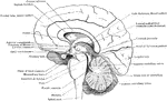
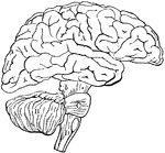
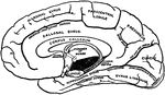
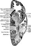
Brain in Mesial Section
Simplified drawing of brain as seen in mesial section, showing relation of brain stem, cerebrum and…
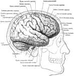
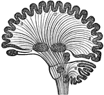
Diagram of Human Brain in Vertical Section
Diagram of human brain in vertical section, showing the situation of the different ganglia and the course…
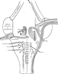
Connection of Brain Nerves
Diagram showing the brain connections of the vagus, glossopharyngeal, auditory, facial, abducent, and…
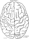
Brain Seen from Above
Labels: 1, longitudinal fissure separating the hemispheres; 2, frontal lobes of the cerebrum; 3, posterior…
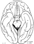
Brain Seen from Below
Labels: 1, longitudinal fissure separating the hemispheres; 2 and 3, front and posterior lobes of the…
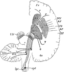
Brain Showing Connection of Frontal Occipital Lobe with Cerebellum
Diagram to show the connecting of the Frontal Occipital Lobes with the Cerebellum. The dotted lines…
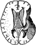
Brain Showing Corpus Callosum
Shown is the corpus callosum. The corpus callosum is a thick stratum of transversely directed nerve…
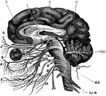
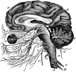
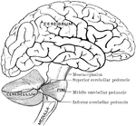
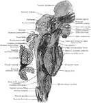
Brain Stem and Adjacent Structures
The right lateral aspect of the brain stem after the cerebral hemisphere (except the Corpus striatum)…
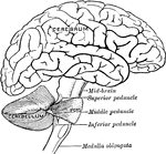
Relation of Brain Stem to Spinal Cord
Simplified drawing of brain as seen from below, showing relations of brain stem to spinal cord and cerebrum.
Sagittal Section of Brain Stem
Sagittal section of brain stem; plane of section is somewhat lateral to midline.
Brain Viewed from Above
The brain viewed from above. Labels: 1, occipital convolution; 2, occipital lobe; 3, inner parietal…
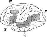
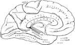
Association Area of the Brain
Lateral view of a brain hemisphere; cortical area V, damage to which produces "mind blindness" (word…
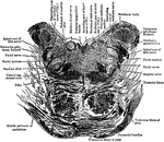
Base of Brain
The base of the brain. Labels: 1, longitudinal fissure; 2, 2, anterior lobes of cerebrum; 3, olfactory…
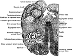
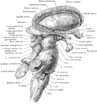
The Base of the Brain
The base of the brain. The cerebral hemispheres are seen overlapping all the rest. Labels: I, olfactory…
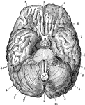
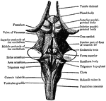
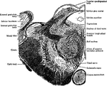
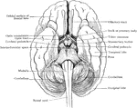
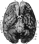
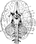
Base of the Brain
The base of brain. Labels: 1. Olfactory Bulb; 2. Second, or Optic Nerves; 3. Anterior Perforated Space;…