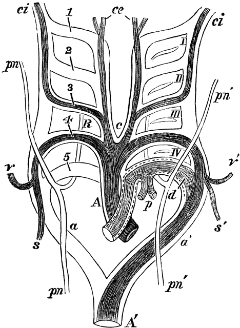
Aortic Arches in a Mammal

Diagram of the aortic arches in a mammal, showing transformations which give rise to the permanent arterial vessels. A, primitive arterial stem or aortic bulb, now divided into A, the ascending part of the aortic arch, and p, the pulmonary; a, a’, right and left aortic roots; A’, descending aorta; 1, 2, 3, 4, 5, the five primitive aortic or branchial arches; I, II, III, IV, the four branchial clefts which, for the sake of clearness, have been omitted on the right side. The permanent systemic vessels are deeply, the pulmonary arteries lightly, shaded; the parts of the primitive arches which are transitory are simply outlined; c, placed between the permanent common carotid arteries; c e, external carotid arteries; c i, internal carotid arteries; s, right subclavian, rising from the right aortic root beyond the fifth arch; v, right vertebral from the same, opposite the fourth arch; v’ s’, left vertebral and subclavian arteries rising together from the left, or permanent aortic root, opposite the forth arch; p, pulmonary arteries rising together from the left fifth arch, forming ductus arteriosus; p n, p n’, right and left pneumogastric nerves descending in from of aortic arch, with their recurrent branches represented diagrammatically as passing behind, to illustrate the relations of these nerves respectively to the right subclavian artery (4) and the arch of the aorta and ductus arteriosus (d).
Galleries
Mammal Anatomy: Internal OrgansSource
Baker, W. Morrant & Harris, Vincent Dormer Kirkes' Hand-book of Physiology, 13th ed. (Philadelphia: P. Blakiston's Son & Co., 1892) 825
Downloads
1758×2400, 1.0 MiB
750×1024, 147.1 KiB
468×640, 72.4 KiB
234×320, 23.7 KiB