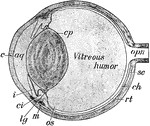
The Mammal Anatomy: Internal Organs ClipArt gallery offers 192 views of the internal organs and other soft tissue of various species of mammals.
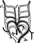
Aortic Arches
Aortic arches of a mammal, and their relations to the five embryonic arches. Labels: c, c', carotids;…
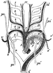
Aortic Arches in a Mammal
Diagram of the aortic arches in a mammal, showing transformations which give rise to the permanent arterial…
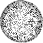
Blood Vessels of the Capsulopupillary of a Kitten
Blood vessels of the capsulopupillary membrane of a newborn kitten.
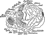
Motor Area of the Brain of a Chimpanzee
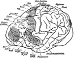
Location of the motor areas in the brain of a chimpanzee. The extent of the motor area is indicated…

Brain of a Dog
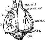
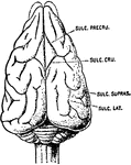
Brain of dog, viewed from above and in profile. F, frontal fissure sometimes termed crucial sulcus,…
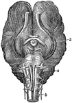
Base of Brain of a Horse
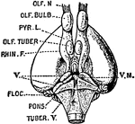
The base of brain of a horse. Labels: l, Cerebrum. 2, Ganglion of sight. 3, Cerebellum. 4, Medulla Oblongata…

Brain of a Monkey to Show effects of Electric Stimulation
Diagrams of monkey's brain to show the effects of electric stimulation of certain spots. Labels: 1.…

Cerebellum of Dog's Brain
Vertical section of dog's cerebellum. Labels: p m, pia mater; p, corpuscles of Purkinje, which are branched…

Horizontal Section of a Vertebrate Brain
Diagrammatic horizontal section of a vertebrate brain. Mb, midbrain: what lies in front of this is the…
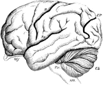
Brain of the Orangutan
Brain of the Orangoutang, showing arrangement of the convolutions. Sy, fissure of Sylvius; R, fissure…

Vertical Section of a Vertebrate Brain
Longitudinal and vertical diagrammatic section of a vertebrate brain. Mb, midbrain: what lies in front…
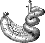
Caecum and Colon of a Dog
Caecum and colon of a dog-inflated. Labels: a, ileum; b, caecum; c, colon.
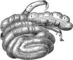
Caecum and Colon of a Hog
Caecum and colon of a hog-inflated. Labels: a, ileum; b, caecum; c, colon; d, rectum.
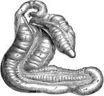
Caecum and Colon of Horse
Caecum and great colon of a horse. Labels: a, caecum; b, c, its muscular bands; d, termination of the…
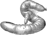
Caecum of an Ox
Caecum and origin of colon of an ox- inflated. Labels: a, terminal portion of the ileum; b, caecum;…
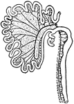
Intestinal Tract from Canis Vulpes
Intestinal tract of Canis vulpes. S, cut end of duodenum; C, caecum; R, cut end of rectum.
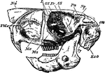
Cape Jumping Hare
"Side view of skull of Cape Jumping Hare. Pmx, premaxilla; Mx, maxilla; Ma, malar; Fr, frontal; L, lachrymal;…

Capillaries from Vitreous Humour of Calf
Capillaries from the vitreous humor of a fetal calf. Two vessels are seen connected by a cord of protoplasm,…
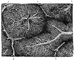
Capillary Network in the Lobules of a Rabbit's Liver
The liver is made up of small roundish or oval portions called lobules, each of which is composed of…

Castor Fiber
"Vertical and Longitudinal section through skull of Castor Fiber, showing the cerebral cavity, the greatly-developed…
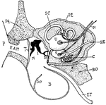
Cat Ear
"Diagram showing the ear and related parts in a young cat. P., Pinna; Sq., squamosal: E.A.M., external…
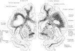
Coronal Sections of the Cerebral Hemispheres
Two coronal sections through the cerebral hemisphere of an orangoutang, in the plan of the anterior…
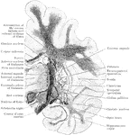
Coronal Section Through Cerebrum
Coronal section through the cerebrum of an orangoutang passing through the subthalamic tegmental region.
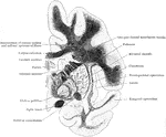
Coronal Section Through the Cerebrum
Coronal section through the left side of the cerebrum of an orangoutang. The section passes through…

Chimpanzee Brain
"Brain of chimpanzee. Ol, olfactory lobe; A, B, C, frontal, occipital, and temporal lobes; C1, a portion…

Lamella of Kitten's Cornea
Surface view of part lamella of kitten's cornea, prepared first with caustic potash and then with nitrate…

Magnified Rabbit's Cornea
Vertical section of rabbit's cornea. Labels: anterior epithelium, showing the different shapes of the…

Section of Rabbit's Cornea
Vertical section of rabbit's cornea, stained with gold chloride. Labels: e, Laminated anterior epithelium.…

Corti from the Dog
Vertical section of the organ of Corti from the dog. Labels: 1 to 2, homogeneous layer of the so-called…
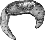
Cow Fetus
Fetus of a cow, with its membrane. Labels: a, placenta; b, chorion with the allantois adherent to its…

Heart of a Deer
"Cast or mold of the interior of the left ventricle of the heart of a deer. Shows that the left ventricular…
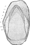
Dental Sac and Pulp
Vertical transverse section of the dental sac and pulp of a kitten. Labels: a, dental papilla or pulp;…
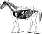
Digestive Apparatus of the Horse
The digestive apparatus of the horse. Labels: a, mouth; 2, pharynx; 3, esophagus; 4, diaphragm; 5, spleen;…

Digestive Organs of a Dog
Stomach, liver, pancreas, and duodenum of a dog. Labels: a, liver; b, gall bladder; c, biliary canals;…

Digestive Organs of a Horse
The relation of anterior abdominal digestive organs- left antero-lateral view. Labels: 1, liver; 2,…

Dog Fetus
Fetus of a dog, with its membrane. Labels: a, placenta; b, chorion with the allantois adherent to its…
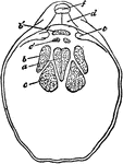
Dorsal Valve
"Waldheimia flavescens. Interior of dorsal valve. c, c', cardial process; b', hinge-plate; s, dental…
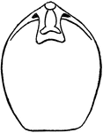
Dorsal Valve
"Terebratula virea. Interior of dorsal valve. l, loop; b, hinge-plate; c, cardinal process." —…
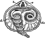
Dorsal Valve
"Rhynchonella psittacea. Interior of doral valve. s, sockets; b, dental plates; V, mouth; de, labial…

Ligaments of the Elbow Joint
The ligaments of the elbow joint- posterior view. Labels: a, external lateral ligament; b, internal…

Epididymis of a Dog
Section of the epididymis of a dog. The tube is cut in several places, both transversely and obliquely,…

Eye Nerves of a Horse
Right orbit opened to show the nerves of the eye. Labels: a, optic; b, motor oculi; c, pathetic; d,…
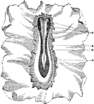
Germinal Membrane of a Dog
Portion of the germinal membrane, with rudiments of the embryo, from the ovum of a dog. The primitive…
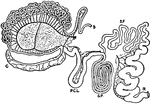
Intestinal Tract of Giraffe
S, cut end of duodenum; R, cut end of rectum; C, caecum; P.C.L., post-caecal loop; S.P., spiral loop;…
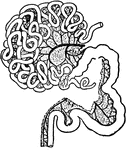
Intestinal Tract of a Gorilla
S, cut end of duodenum; R, cut end of rectum; C, vermiform appendix of caecum; X1, X2, X3, cut ends…
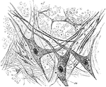
Gray Matter of Spinal Cord
Section of gray matter of anterior cornu of a calf's spinal cord; a, nerve fibers of white matter in…

Greenland Whale
This illustration shows the jaw of a Greenland Whale. The Greenland Whale uses this massive jaw to filter…
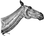
Head and Neck of a Horse Showing Veins
Veins of the face and neck. Labels: 1, glosso-facial; A, its facial portion; 2, jugular; 3, occipial;…

Head of a Horse
Longitudinal section of the head, showing the pharynx and nasal chamber-the septum nasi being removed.…

Head of a Horse Showing Arteries
Arteries of the head- the left maxillary ramus being remove. Labels: 1, occipital; 2, internal carotid;…
