The Mammal Anatomy: Internal Organs ClipArt gallery offers 192 views of the internal organs and other soft tissue of various species of mammals.

Tendons and ligaments of the left anterior extremity of ox, viewed from external side. Labels: a, flexor perforatus tendon; a', its superficial; a", its deep branch; b, interosseus metacarpeus; c, flexor perforans tendon; d-d", e-e"', superior suspensory ligament and attachments; d, anterior division; e', e", branches forming superior ligaments of small claw; e'". inferior ligament of small claw; f, common annular ligament; g, g', superior and inferior special annular ligaments; h, interdigital ligament; i, i', tendon of the extensor proprius externus.

Tendons and Ligaments of Ox Leg

Tendons and ligaments of the left anterior extremity of ox, viewed from external side. Labels: a, flexor…

From a section of the testis of a dog, showing portions of seminal tubes. A, seminal epithelial cells, and numerous small cells loosely arranged; B, the small cells or spermatoblasts converted into spermatozoa; groups of these is a further stage of development.

Testis of a Dog, Section of the

From a section of the testis of a dog, showing portions of seminal tubes. A, seminal epithelial cells,…

Internal view of left thigh-showing the arteries. Labels: 1, femoral; a, profunda femoris; b, superficialis femoris; c, saphenic; d, muscular branches; e, femora; nutrient vessels cut; (the femoral becomes the popliteal at about this point); f, femoro-popiteal; f', its muscular branches; f", its descending branch; g, recurrent tibial; h, united trunk of saphenic and femoro-popliteal; i, internal plantar.

Thigh of a Horse Showing Arteries

Internal view of left thigh-showing the arteries. Labels: 1, femoral; a, profunda femoris; b, superficialis…
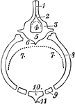
Diagrammatic transverse section from the skeleton of a mammalian thorax, showing the chief features of a perfect vertebral segment. 1, neural spine; 2, lamina (neurapophysis); 3, [edicle; 4, neural arch; 5, centrum; 6, inferior spine (hypapophysis); 7, haemal arch; 8, rib (pleurapophysis); 9, costal cartilage (haemapophysis); 10, sternebra; 11, haemal spine.

Diagram of Mammilian Thorax

Diagrammatic transverse section from the skeleton of a mammalian thorax, showing the chief features…
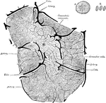
Minute structure of the thymus gland. Lobule of inject thymus from a calf, four days old, slightly diagrammatic, magnified about 50 diameters. The large vessels are disposed in two rings, one of which surrounds the lobule, the other lies just within the margin of the medulla.

Structure of the Thymus

Minute structure of the thymus gland. Lobule of inject thymus from a calf, four days old, slightly diagrammatic,…
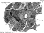
Minute structure of the thyroid. From a transverse section of the thyroid of a dog.

Section of Dog's Thyroid Gland

Minute structure of the thyroid. From a transverse section of the thyroid of a dog.
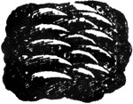
"Felidæ or Felinæ is the cat tribe, a family of carnivorous quadrupeds, including the domestic cat, lions, tigers, panthers, leopards, and lynxes. The felidæ are found in all parts of the world except Australia. "—(Charles Leonard-Stuart, 1911)

Partial Tiger Tongue

"Felidæ or Felinæ is the cat tribe, a family of carnivorous quadrupeds, including the domestic…

A, Section through papilla vallata of a human tongue. B, Section through part of the papilla foliata of a rabbit. Labels: 1, papilla; 2, vallum; 3, taste buds; 4, papillae; 5, taste buds; 6, ducts of serous gland.

Tongue of Human and Rabbit

A, Section through papilla vallata of a human tongue. B, Section through part of the papilla foliata…
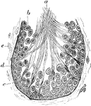
Section of a tubule of the testicle of a rat, to show the formation of the spermatozoa. Labels: a, spermatozoa; b, seminal cells; c, spermatoblasts, to which the spermatozoa are sill adherent; d, membrana propria; e, fibro-plastic elements of the connective tissue.

Section of a Tubule of the Testicle of a Rat

Section of a tubule of the testicle of a rat, to show the formation of the spermatozoa. Labels: a, spermatozoa;…

"Longitudinal section of valves. A, ventral, B, dorsal valves; l, loop; q, crura; ss, septum; c, cardinal process." — Encyclopedia Britanica, 1893

Valves

"Longitudinal section of valves. A, ventral, B, dorsal valves; l, loop; q, crura; ss, septum; c, cardinal…
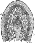
Section of the villus of a rat killed during fat absorption. Labels: ep, epithelium; str, striated border; c, lymph cells; c', lymph cells in the epithelium; l, central lacteal containing disintegrating lymph corpuscles

Villus of a Rat

Section of the villus of a rat killed during fat absorption. Labels: ep, epithelium; str, striated border;…
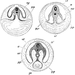
Diagram showing the three successive stages of development. Transverse vertical sections. The yolk sac, ys, is seen progressively diminishing in size. In the embryo itself the medullary canal and notochord are seen in section. a', in middle figure, the alimentary canal, becoming pinched off, as it were, from the yolk sac; a', in right hand figure, alimentary canal completely closed; a, in last two figures amnion; ac', cavity of amnion filled with amniotic fluid; p, space between amnion and chorion continuous with the pleuroperitoneal cavity inside the body; vt, vitelline membrane; ys, yolk sac, or umbilical vesicle.

Development of the Yolk Sac

Diagram showing the three successive stages of development. Transverse vertical sections. The yolk sac,…