Development of the Yolk Sac

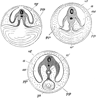
Diagram showing the three successive stages of development. Transverse vertical sections. The yolk sac, ys, is seen progressively diminishing in size. In the embryo itself the medullary canal and notochord are seen in section. a’, in middle figure, the alimentary canal, becoming pinched off, as it were, from the yolk sac; a’, in right hand figure, alimentary canal completely closed; a, in last two figures amnion; ac’, cavity of amnion filled with amniotic fluid; p, space between amnion and chorion continuous with the pleuroperitoneal cavity inside the body; vt, vitelline membrane; ys, yolk sac, or umbilical vesicle.
Galleries
Mammal Anatomy: Internal OrgansSource
Baker, W. Morrant & Harris, Vincent Dormer Kirkes' Hand-book of Physiology, 13th ed. (Philadelphia: P. Blakiston's Son & Co., 1892) 803
Downloads
2384×2400, 710.7 KiB
1017×1024, 114.1 KiB
635×640, 59.9 KiB
317×320, 21.5 KiB