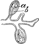
"A, female gonophores of Athorybia rosacea on their common stem or gynophore: a, ovum; b, radial canals." -Whitney, 1911

Athorybia Gonophores

"A, female gonophores of Athorybia rosacea on their common stem or gynophore: a, ovum; b, radial canals."…

Every human body begin as a single nucleated cell. This cell, known as the ovum, divides or segments and gives rise to a mass consisting of a number of similar units known as the morula. At this stage there are no distinguishable tissues.

Cell Development

Every human body begin as a single nucleated cell. This cell, known as the ovum, divides or segments…

Very soon after the entrance of the ovum into the uterus, in the human subject, the outer surface of the chorion is found beset with fine processes, the so-called chorion villi, which give it a rough and shaggy appearance. a, chorion with villi. The villi are shown to be best developed in the part of the chorion to which the allantois is extending, this portion ultimately becomes the placenta. ; b, space between the two layers of the amnion; c, amniotic cavity; d, situation of the intestine, showing its connection with the umbilical vesicle; e, umbilical vesicle; f, situation of heart and vessels; g, allantois.

Chorion Villi

Very soon after the entrance of the ovum into the uterus, in the human subject, the outer surface of…
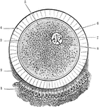
The ovum and its coverings. The corona radiata, which completely surrounds the ovum, is only represented in the lower part of the figure. Labels: 1, corona radiate; 2, granular layer; 3, vitelline membrane; 4, zona pellucida (oolemma); 5, vitellus or yolk; 6, germinal vesicle (nucleus); 7, germinal spot (nucleolus); 8, nuclear membrane.

Ovum

The ovum and its coverings. The corona radiata, which completely surrounds the ovum, is only represented…

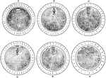
Sectional views of the ovum, after segmentation.

Ovum After Segmentation

Sectional views of the ovum, after segmentation.

Diagram of a developing ovum, seen in longitudinal section. Labels: a, pericardium; b, bucco-pharyngeal; c, ectoderm; d, entoderm; e,placental area. f, spinal cord; i, brain; g, notochord; k, extra embryonic coelom; h, dorsal wall of alimentary canal.

Developing Ovum

Diagram of a developing ovum, seen in longitudinal section. Labels: a, pericardium; b, bucco-pharyngeal;…
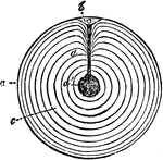
"Meroblastic ovum (yelk) of domestic fowl, bat. size, in section; after haeckel. a, the thin yelk-skin, enclosing the yellowfood-yelk, which is deposited in concentric layers, c, d; b, the cicatricle or tread with its nuclues, whence passes a cord of white yelk (here represented in black) to the central cavity, d'" Elliot Coues, 1884

Fowl Ovum

"Meroblastic ovum (yelk) of domestic fowl, bat. size, in section; after haeckel. a, the thin yelk-skin,…
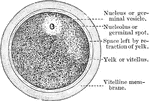
The diagram of a human ovum, showing the parts of an animal cell.

A Diagram of the Human Ovum

The diagram of a human ovum, showing the parts of an animal cell.

The maturation of the ovum. A, An ovum at the commencement process. B, After the formation of the spindle. The chromosomes are gathered at the equator of the spindle in groups of four, i.e. in tetrads, each which consists of two dyads. C, One apex of the spindle has projected into a bud on the surface, and the dyads have passed to the poles. D, The separation of the first polar body. E, The commencement of the second polar body; F, The completion of the second polar body.

Maturation of the Ovum

The maturation of the ovum. A, An ovum at the commencement process. B, After the formation of the spindle.…
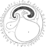
Diagrammatic section showing the relation in a mammal between the primitive alimentary canal and the membranes of the ovum. The stage represented in this diagram corresponds to that of the fifteenth or seventeenth day in the human embryo, previous to the expansion of the allantois; c, the villous chorion; a, the amnion; a', the place of convergence of the amnion and reflexion of the false amnion a", or outer or corneous layer; e, the head and trunk of the embryo, comprising the primitive vertebrae and cerebrospinal axis; i, the simple alimentary canal in its upper and lower portions. Immediately beneath the right hand i is seen the fetal heart, lying in the anterior part of the pleuroperitoneal cavity; v, the yolk sac or umbilical vesicle; vi, the vitellointestinal opening; u, the allantois connected by a pedicle with the anal portion of the alimentary canal.

Membranes of the Ovum

Diagrammatic section showing the relation in a mammal between the primitive alimentary canal and the…
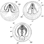
Diagram showing the three successive stages of development. Transverse vertical sections. The yolk sac, ys, is seen progressively diminishing in size. In the embryo itself the medullary canal and notochord are seen in section. a', in middle figure, the alimentary canal, becoming pinched off, as it were, from the yolk sac; a', in right hand figure, alimentary canal completely closed; a, in last two figures amnion; ac', cavity of amnion filled with amniotic fluid; p, space between amnion and chorion continuous with the pleuroperitoneal cavity inside the body; vt, vitelline membrane; ys, yolk sac, or umbilical vesicle.

Development of the Yolk Sac

Diagram showing the three successive stages of development. Transverse vertical sections. The yolk sac,…