The Mammal Anatomy: Internal Organs ClipArt gallery offers 192 views of the internal organs and other soft tissue of various species of mammals.

"Thumb and leg and foot of Mystacina tuberculata." —The Encyclopedia Britannica, 1903

Mystacina Tuberculata

"Thumb and leg and foot of Mystacina tuberculata." —The Encyclopedia Britannica, 1903

Navicular ligaments. Labels: a, superior, or broad, ligament; b, inferior ligament; c, lateral ligament.

Navicular Ligaments

Navicular ligaments. Labels: a, superior, or broad, ligament; b, inferior ligament; c, lateral ligament.

Arteries of the neck exposed on the left side. Labels: a, anterior aorta; a', left brachial; a", right brachial, or bracio-cephalic; b, left dorsal; c, superior cervical; d, vertebral; e, inferior cervical; f, cephalic or common carotid; f", left , and f", right carotid; g, esophageal wigs; h, tracheal twigs; i, muscular twigs; k, thyroid; l, thyro-laryngeal; m, branch to parotid gland.

Neck of a Horse Showing Arteries

Arteries of the neck exposed on the left side. Labels: a, anterior aorta; a', left brachial; a",…
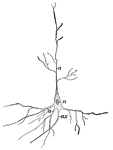
Nerve cell from the brain; <em>a</em>, processes by which it communicates with other cells near by; <em>ax</em>, long process or axis cylinder terminating in the tail end of the spinal cord; <em>n</em>, nucleus.

Nerve Cell

Nerve cell from the brain; a, processes by which it communicates with other cells near by;…
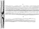
Gray, pale, or gelatinous nerve fibers. A. From a branch of olfactory nerve of the sheep: two dark bordered or white fibers from the fifth pair are associated with the pale olfactory fibers. B. From the sympathetic nerve.

Gelatinous Nerve Fibers

Gray, pale, or gelatinous nerve fibers. A. From a branch of olfactory nerve of the sheep: two dark bordered…

Each nerve trunk is composed of a variable number of different sized bundle (funiculi) of nerve fibers which have a special sheath (perineurium) The funiculi are enclosed in a firm fibrous sheath (epineurium); this sheath also send in processes of connective tissue which connect the bundles together. In the funiculi between the fibers is a delicate supporting tissue ( the endoneurium). Shown is a transverse section of the sciatic nerve of a cat. It consists of bundles (Funiculi) of nerve fibers ensheathed in a fibrous supporting capsule, epineurium, A; each bundle has a special sheath ( not sufficiently marked out from the epineurium in the figure) or perineurium B; the nerve fibers N f are separated from one another by endoneurium; L, lymph spaces; Ar, artery; V, vein; F, fat.

Nerve Trunk

Each nerve trunk is composed of a variable number of different sized bundle (funiculi) of nerve fibers…
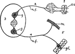
"Scheme showing the essential relations of the parts of a nervous system: 1, the sensory end organ (epithelial); 2, afferent nerve tract; 3, central nervous cells (ganglia); 4, efferent nerves, leading to 5, muscle, gland, etc. g, ganglion cells; gl., gland; m, muscle fibre; n.f., nerve fibre; s.e., sensory epithelium." &mdash; Galloway

Nervous System

"Scheme showing the essential relations of the parts of a nervous system: 1, the sensory end organ (epithelial);…
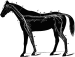
The nervous system of the horse. Labels: 1, brain; 2, optic nerve; 3, superior maxillary nerve (5th); 4, inferior maxillary nerve (5th); 5, pneumogastric nerve; 6, medulla oblongata; 6', spinal cord; 7, right brachial plexus; 8, musculo-cutaneous nerve; 9, median nerve; 10, radial nerve; 11, cubital nerve; 12, pneumogastric nerve; 13, solar plexus; 14, semilunar ganglion; 15m lumbo-sacral plexus; 16, anterior crural nerve; 17, great sciatic nerve; 18, external popiteal nerve; 19, external saphenic nerve; 20, tibial nerve; 21, metatarsal nerve; 22, radial portion of median nerve; 23, metacarpal nerves; 24, digital branches; s, sympathetic chain; c, inferior cervical plexus; g, gutteral ganglion; Sp, great splanchnic nerve; pm; posterior mesenteric plexus; p, pelvic plexus.

Nervous System of the Horse

The nervous system of the horse. Labels: 1, brain; 2, optic nerve; 3, superior maxillary nerve (5th);…

The neural canal of first three cervical vertebrae, opened from above to show the internal ligaments. The occiput and foramen magnum are seen to the right. Labels: b, the odontoid ligament; a, the occipitoatloid portion (long adontoid); c, superior common ligament; d, posterior articular cavity of a vertebral centrum.

Neural Canal

The neural canal of first three cervical vertebrae, opened from above to show the internal ligaments.…

"The size of the hemispheres of the brain (A) is so small that they leave exposed the olfactory ganglion (a), the cerebellum (C), and more or less of the optic lobes (B), and they are but partially connected together by the 'fornix' and 'anterior commissure,' the great cerebral commissure known as the 'corpus callosum' being absent." &mdash; Chambers, 1881

Opossum Brain

"The size of the hemispheres of the brain (A) is so small that they leave exposed the olfactory ganglion…
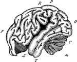
"The outline of the brain of an orang outang. Front portion F to O, cerebrum; C, cerebellum; M, medulla and spinal cord; F, the frontal lobe; P, the parietal lobe; O, the occipital lobe; T, the temporal lobe; R, the fissure of Rolando; S, the fissure of Silvius." -Galloway, 1915

Orangoutang Brain

"The outline of the brain of an orang outang. Front portion F to O, cerebrum; C, cerebellum; M, medulla…

Diagram of the chief organs of a mammal. The bones are black. <em>a</em>, opening from the nasal cavity <em>s</em> to pharynx; <em>bl</em>, bladder; <em>ce</em>, caecum; <em>di</em>, diaphragm; <em>e</em>, epiglottis; <em>n</em>, nostril or anterior nares; <em>ov</em>, ovary; <em>rf</em> and <em>ute</em>, uterus; <em>ru</em> and <em>lu</em>, ureters; <em>x</em>, pancreas; <em>vag</em>, vagina. From Davison's <em>Mammalian Anatomy</em>.

Organs

Diagram of the chief organs of a mammal. The bones are black. a, opening from the nasal cavity…
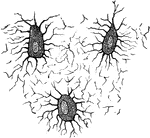
Nucleated none cells (osteoblasts) and their processes, contained in the one lacunae and their canaliculi respectively. From a section through the vertebra of an adult mouse.

Osteoblasts

Nucleated none cells (osteoblasts) and their processes, contained in the one lacunae and their canaliculi…
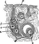
"Section through ovary of a young Mammal. The eggs (o) are seen to be formed from the epithelium. c, connective tissue of ovary; e, epithelium; f, follicle of epithelial cells in which the ova ripen; o, ova in different stages of ripeness." &mdash; Galloway

Ovary

"Section through ovary of a young Mammal. The eggs (o) are seen to be formed from the epithelium. c,…
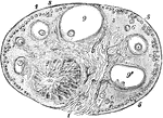
View of a section of the ovary of the cat. Labels: 1, outer covering and free border of the ovary; 1', attached border; 2, the ovarian stroma, presenting a fibrous and vascular structure; 3, granular substance lying external to the fibrous stroma; 4, blood-vessels; 5, ovigerms in their earliest stages occupying part of the granular layer near the surface; 6, ovigerms which have begun to enlarge and to pass more deeply into the ovary; 7, ovigerms round which the Graafian follicles and tunica granulosa are now formed and which have passed somewhat deeper into the ovary and are surrounded by the fibrous stroma; 8, more advanced Graafian follicle with the ovum imbedded in the layer of cells constituting the proligerous disc; 9, the most advanced follicle containing the ovum; 9', a follicle from which the ovum has accidentally escaped; IO, corpus luteum.

Ovary of the Cat

View of a section of the ovary of the cat. Labels: 1, outer covering and free border of the ovary; 1',…
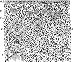
Section of the ovary of a cat. Labels: A, germinal epithelium; B, immature Graafian follicle; C, stroma of ovary; D, vitelline membrane containing the ovum; E, Graafian follicle showing lining cells; F, follicle from which the ovum has fallen out.

Ovary of the Cat

Section of the ovary of a cat. Labels: A, germinal epithelium; B, immature Graafian follicle; C, stroma…
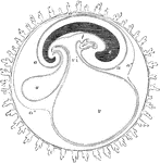
Diagrammatic section showing the relation in a mammal between the primitive alimentary canal and the membranes of the ovum. The stage represented in this diagram corresponds to that of the fifteenth or seventeenth day in the human embryo, previous to the expansion of the allantois; c, the villous chorion; a, the amnion; a', the place of convergence of the amnion and reflexion of the false amnion a", or outer or corneous layer; e, the head and trunk of the embryo, comprising the primitive vertebrae and cerebrospinal axis; i, the simple alimentary canal in its upper and lower portions. Immediately beneath the right hand i is seen the fetal heart, lying in the anterior part of the pleuroperitoneal cavity; v, the yolk sac or umbilical vesicle; vi, the vitellointestinal opening; u, the allantois connected by a pedicle with the anal portion of the alimentary canal.

Membranes of the Ovum

Diagrammatic section showing the relation in a mammal between the primitive alimentary canal and the…
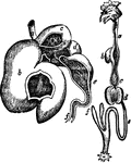
"Compound stomach of ox. a, esophagus; b, rumen, or paunch; c, reticulum, or second stomach; d, omasum, or third stomach; e, abomasum, or fourth stomach; f, the duodenuin." &mdash; Chambers' Encyclopedia, 1875

Ox Stomach

"Compound stomach of ox. a, esophagus; b, rumen, or paunch; c, reticulum, or second stomach; d, omasum,…
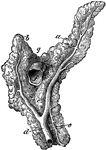
Anterior view of the pancreas. Labels: a, left branch; b, right branch; c, inferior branch; d, duct of Wirsung; e, ductus pancreaticus minor; f, portal vein cut across; g, notch for the great mesenteric artery.

Pancreas of a Horse

Anterior view of the pancreas. Labels: a, left branch; b, right branch; c, inferior branch; d, duct…
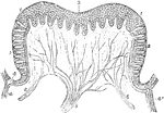
Vertical section of a circumvallate papilla of the calf. Labels: 1 and 3, epithelial layers covering it; 2, taste goblets; 4 and 4', duct of serous gland opening out into the pit in which papilla is situated; 5 and 6, nerves ramifying within the papilla.

Papilla of a Calf

Vertical section of a circumvallate papilla of the calf. Labels: 1 and 3, epithelial layers covering…

Parotid and molar glands of the left side. Labels: a, parotid gland; b, Steno's duct; c, superior molar glands; d, inferior molar gland.

Parotid and Molar Glands of a Horse

Parotid and molar glands of the left side. Labels: a, parotid gland; b, Steno's duct; c, superior molar…
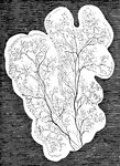
In mammalia, each salivary gland first appears as a simple canal with bud-like processes, lying in a gelatinous nidus or blastema, and communicating with the cavity of the mouth. As the development of the gland advances, the canal becomes more and more ramified, increasing at the expense of the blastema in which it is still enclosed. the branches or salivary ducts constitute an independent system of closed tubes. Shown are lobules of the parotid, with salivary ducts, in the embryo of the sheep at a more advanced stage.

Lobules of Parotid of a Sheep

In mammalia, each salivary gland first appears as a simple canal with bud-like processes, lying in a…
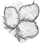
The glands of Peyer occur chiefly but not exclusively in the small intestine. They are found in greatest abundance in the lower part of the ileum near to the ileocaecal valve. Shown is a transverse section of injected Peyer's glands representing the fine capillary-looped network spreading from the surrounding blood vessels into the interior of three of Peyer's capsules from the intestine of the rabbit.

Peyer's Glands

The glands of Peyer occur chiefly but not exclusively in the small intestine. They are found in greatest…

"Brain of pig. Ol, olfactory lobe; A, B, C, frontal, occipital, and temporal lobes; C1, a portion of temporal lobe; Sy, Sylvian fissure; In, insula or island of Reil." -Whitney, 1911

Pig Brain

"Brain of pig. Ol, olfactory lobe; A, B, C, frontal, occipital, and temporal lobes; C1, a portion of…
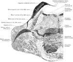
Section through the pons varolii at the level of the nuclei of the trigeminal nerve in an orangoutang.

Section Through Pons Varolii

Section through the pons varolii at the level of the nuclei of the trigeminal nerve in an orangoutang.

Section through the upper part of the pons varolii of the orangoutang above the level of the trigeminal nerve.

Section Through Pons Varolii

Section through the upper part of the pons varolii of the orangoutang above the level of the trigeminal…
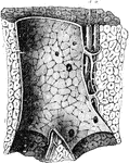
Longitudinal section of a portal canal, containing a portal vein, hepatic artery and hepatic duct from the pig. Labels: P, branch of vena portae, situate in a portal canal formed amongst the lobule of the liver; l, and giving off vaginal branches; there are also seen within the larger portal vein numerous orifices of the small interlobular veins arising directly from it; a, hepatic artery; d, hepatic duct.

Portal Canal

Longitudinal section of a portal canal, containing a portal vein, hepatic artery and hepatic duct from…

the internal anatomy of a rabbit.

Rabbit

the internal anatomy of a rabbit.

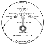
Cross section of abdomen of a rabbit.

Rabbit

Cross section of abdomen of a rabbit.

"Brain of rabbit. Ol, olfactory lobe; A, B, C, frontal, occipital, and temporal lobes; Sy, Sylvian fissure." -Whitney, 1911

Rabbit Brain

"Brain of rabbit. Ol, olfactory lobe; A, B, C, frontal, occipital, and temporal lobes; Sy, Sylvian fissure."…
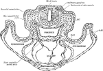
Transverse section of a rat embryo. Showing relation of the paraxial mesoderm of the head to the lateral plates, the commencement of the formation of the otic vesicles and hyomandibular clefts, and the relation of the primitive heart to the pericardium and foregut. Labels: EC., ectoderm; SoM., somatic mesoderm; SpM., splanchnic mesoderm.

Transverse Section of a Rat Embryo

Transverse section of a rat embryo. Showing relation of the paraxial mesoderm of the head to the lateral…

General view of the respiratory organs. Labels: a, septum nasi; b, posterior naris; c, larynx; d, trachea; e, thyroid gland; f, anterior mediastinum; g, posterior mediastinum; h, left lung; h', its apex; h&quot;, its base; h&quot;', its superior,and h&quot;&quot;, its inferior borders.

Respiratory Organs of a Horse

General view of the respiratory organs. Labels: a, septum nasi; b, posterior naris; c, larynx; d, trachea;…

The respiratory apparatus of other mammals is similar to humans in both structure and function. The lungs fill the cavity of the chest, and have the same general composition of lobes, lobules and air-cells. Labels: 1, Trachea. 2, Lungs. 3, Heart. 4, Diaphragm. 5, Liver. 6, Stomach. 7, Esophagus. 8, Kidney. 9, Intestines. 10, Bladder. 11, Cerebrum. 12, Cerebellum. 13, Medulla spinalis. 14, Vertebrae.

The Respiratory System of a Small Mammal

The respiratory apparatus of other mammals is similar to humans in both structure and function. The…

Perpendicular section of mammalian retina. Labels: A, layer of rods and cones; B, outer nuclear layer; C, outer molecular layer; D, inner nuclear layer; E, inner molecular layer; F, ganglionic layer; G, stratum opticum; r, rods; c, cones, r.g, rod granules; c.g, cone granules; r.b, rod bipolars; c.b, cone bipolars; c.r, vontact of rod bipolars with the internal arborisation of cone bipolars; ar', internal arborisation of rod bipolars; c.n, centrifugal nerve fiber; h, horizontal cells; s.s, stratified spongioblasts; d.s, diffuse spongioblasts; s.g, stratified ganglion cell; M, sustentacular fiber of Muller.

Section of Retina

Perpendicular section of mammalian retina. Labels: A, layer of rods and cones; B, outer nuclear layer;…
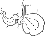
"Diagram of the stomach of a ruminant. o, esophagus; r, rumen or paunch; re., reticulum, or honeycomb; p, psalterium or manyplies; a, abomasum or rennet; i, intestine." -Galloway, 1915

Ruminant Stomach

"Diagram of the stomach of a ruminant. o, esophagus; r, rumen or paunch; re., reticulum, or honeycomb;…

Serous glands. Labels: a, rabbit's pancreas "loaded" (resting); c, "discharged" (active) (observed in the living animal). b, loaded; d, discharged, alveolus of parotis (fresh preparation).

Serous Glands of a Rabbit

Serous glands. Labels: a, rabbit's pancreas "loaded" (resting); c, "discharged" (active) (observed in…

Sesamoidean and digital ligaments- posterior aspect. Labels: a, suspensory ligament; b, external and middle inferior sesamoidean ligaments. The central bundle is the Y, the two lateral ones forming the V ligament; c, annular sesamoidean ligament; d, posterior ligaments of the pastern joint; e, lateral navicular ligament; f, inferior navicular ligament.

Sesamoidean and Digital Ligaments

Sesamoidean and digital ligaments- posterior aspect. Labels: a, suspensory ligament; b, external and…

"Stomach of sheep. a, OEsophagus; c, rumen or paunch; d, reticulum or honeycomb-bag; e, psalterium or many-plies; f, abomasum or reed; b, beginning of duodenum." -Thomson, 1916

Sheep Stomach

"Stomach of sheep. a, OEsophagus; c, rumen or paunch; d, reticulum or honeycomb-bag; e, psalterium or…
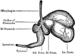
The four stomachs of the sheep, a grass-eating animal. The beginning of the intestines are also shown, which reach in their full length to 28 times the size of the sheep's body.

Sheep, Stomachs of

The four stomachs of the sheep, a grass-eating animal. The beginning of the intestines are also shown,…

Internal aspect of the spleen. Labels: a, superior extremity, or base; b, inferior extremity; c, internal surface; d, the hilus; e, anterior border; f, posterior border; g, suspensory ligament.

Spleen of a Horse

Internal aspect of the spleen. Labels: a, superior extremity, or base; b, inferior extremity; c, internal…

Ligaments of the stifle joint- posterior aspect. Labels: a, external lateral patellar ligament; b, external lateral femoro-tibial ligament; d, external meniscus; e, internal meniscus; f, femoral coronary ligament; g, posterior external coronary ligament; h, fibulo-tibial ligament.

Stifle Joint Ligaments

Ligaments of the stifle joint- posterior aspect. Labels: a, external lateral patellar ligament; b, external…
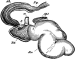
Stomach of a musk deer, left aspect- the last three compartments opened and reflected forwards. Rn, rumen; Rt, reticulum; Ab, abomasum; Py, pylorus; ae, esophagus; X, opening of the tube which represents the psalterium; Spl, spleen.

Stomach of a Deer

Stomach of a musk deer, left aspect- the last three compartments opened and reflected forwards. Rn,…
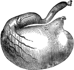
Stomach of a dog-inflated. Labels: a, cardiac portion; b, pyloric portion; c, esophageal orifice; d, duodenum.

Stomach of a Dog

Stomach of a dog-inflated. Labels: a, cardiac portion; b, pyloric portion; c, esophageal orifice; d,…
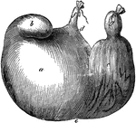
Stomach of a hog- inflated. Labels: a, cardiac portion; b, its accessory cul-de-sac; c, pyloric portion; d, lesser curvature; c, greater curvature; f, esophagus; g, pyloric orifice.

Stomach of a Hog

Stomach of a hog- inflated. Labels: a, cardiac portion; b, its accessory cul-de-sac; c, pyloric portion;…

Stomach of an sheep seen from the left side- the last three compartments are laid open and reflected forwards. Labels: a, esophagus; Ru., rumen; Ret., reticulum; Ps., psalterium; A., abomasum; Du., duodeum. The esophageal canal is seen traversing the lesser curvature of the reticulum.

Stomach of a Sheep

Stomach of an sheep seen from the left side- the last three compartments are laid open and reflected…

Compartments of a ruminant stomach, laid open. A, rumen- a, pillars; b, papilla; c, esophageal orifice. B, reticulum- a, alveoli; b, esophageal canal. C, psalterium- a, hooked papillae; b, valve at orifice leading to abomasum; c, d, e, f, leaves graduated from large to small. D, abomasum- a, folds of mucous membrane; b, pyloric orifice; c, duodenum.

Stomach of a Sheep

Compartments of a ruminant stomach, laid open. A, rumen- a, pillars; b, papilla; c, esophageal orifice.…
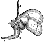
Ruminants (those animals that chew the cud), as the sheep, have a stomach with four cavities. Labels: 1, esophagus; 2, rumen; 3, reticulum; 4, omasum; 5, abomasum or rennet; 6, intestine.

The Stomach of a Sheep

Ruminants (those animals that chew the cud), as the sheep, have a stomach with four cavities. Labels:…
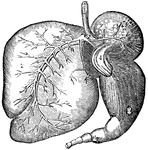
Ruminants (those animals that chew the cud), as the ox, have a stomach with four cavities. Labels: 1, The esophagus. 2, The rumen (paunch). 3, The reticulum (honeycomb). 4, The omasum (many-plies). 5, The abomasum(rennet). 6, The intestine.

Stomach of an Ox

Ruminants (those animals that chew the cud), as the ox, have a stomach with four cavities. Labels: 1,…
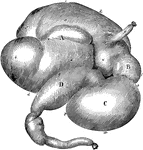
Stomach of an ox inflated-viewed from the right side. Labels: A, rumen; a, left sac; a', its anterior extremity; a&quot;, left conical sac; b, right sac; b', its anterior extremity; b&quot;, right conical sac; f, its dilated extremity entering rumen; h, spleen; B, reticulum; i, its greater curvature; k, its left extremity; l, its right extremity; C, psalterium; m, its greater curvature; n, its lesser curvature; o, its anterior extremity; p, its posterior extremity; D, abomasum; q, its greater curvature; r, its lesser curvature; s, its anterior extremity; t, its posterior extremity; u, pylorus; v, duodenum.

Stomach of an Ox

Stomach of an ox inflated-viewed from the right side. Labels: A, rumen; a, left sac; a', its anterior…
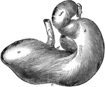
Posterior view of the stomach of a horse. Labels: a, left cul-de-sac; b, right cul-de-sac; c, greater curvature; d, lesser curvature; e, esophagus; f, duodenum.

Horse Stomach

Posterior view of the stomach of a horse. Labels: a, left cul-de-sac; b, right cul-de-sac; c, greater…
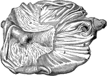
Internal aspect of a horse stomach, opened from below. Labels: a, cuticular mucous membrane; b, villous mucous membrane; c, line of demarcation between the two portions; d, cardiac orifice; e, pyloric orifice and valve.

Horse Stomach

Internal aspect of a horse stomach, opened from below. Labels: a, cuticular mucous membrane; b, villous…
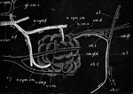
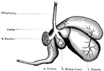
Diagram of a sheep's stomach, showing the different compartments.

Sheep Stomach

Diagram of a sheep's stomach, showing the different compartments.

Diagrammatic representation of the submaxillary gland of the dog with its nerves and blood vessels. Labels: sm. gld., the submaxillary gland into the duct (sm. d.) of which a cannula has been tied. The sublingual gland and duct are not proceeding from the facial nerve, becoming conjoined with the lingual at n.l'., and afterwards diverging and passing to the gland along the duct; sm. gl., submaxillary ganglion with its roots; n. l., the lingual nerve proceeding to the tongue; a. car., the carotid artery. two branches of which, a. sm. a. and r. sm. p. pass to the anterior and posterior parts of the gland; v. sym., the conjoined vagus and sympathetic trunks; gl. cer. s., the superior cervical ganglion, two branches of which forming a plexus, a.f., over the facial artery are distributed (n.sym.sm.) along the two glandular arteries to the anterior and posterior portion of the gland. The arrows indicate the direction taken by the nervous impulses; during reflex stimulations of the gland they ascend to the brain by the lingual and descend by the chorda tympani.

Submaxillary Gland of Dog with Nerves and Blood Vessels

Diagrammatic representation of the submaxillary gland of the dog with its nerves and blood vessels.…
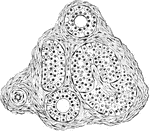
Terminal tubules of sudoriferous glands, cut in various directions from the skin of the pig's ear.

Sudoriferous Glands

Terminal tubules of sudoriferous glands, cut in various directions from the skin of the pig's ear.

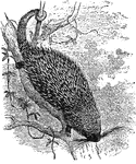
A porcupine found in Mexico and much of South America.

Synetheres Prehensilis

A porcupine found in Mexico and much of South America.

Digestive system of a mammal. (g) gullet; (s) stomach; (sm) small intestine; (lm) large intestine; (r) termination of the large intestine.

Digestive system

Digestive system of a mammal. (g) gullet; (s) stomach; (sm) small intestine; (lm) large intestine; (r)…

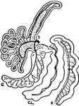
S. cut end of duodenum; R, cut end of rectum; C, caecum; Cl, colon.

Intestinal Tract of a Tapir

S. cut end of duodenum; R, cut end of rectum; C, caecum; Cl, colon.

Isolated cells from taste bud of a rabbit.

Taste bud Cells of a Rabbit

Isolated cells from taste bud of a rabbit.

A, Three-quarter surface view of taste bud from the papilla foliata of a rabbit. B, Vertical section of taste bud from the papilla foliata of a rabbit.

Taste bud of Rabbit

A, Three-quarter surface view of taste bud from the papilla foliata of a rabbit. B, Vertical section…

The sense of taste enables us to test in some degree the chemical constitution of substances we take into the mouth as food. These organs are located in the mouth or on the mouth parts. This example shows a vertical section of large papilla on the tongue of a calf detailing the taste buds

Sense of Taste

The sense of taste enables us to test in some degree the chemical constitution of substances we take…