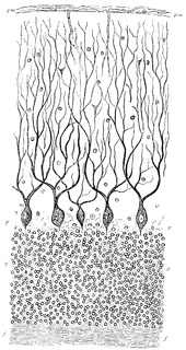
Cerebellum of Dog's Brain

Vertical section of dog’s cerebellum. Labels: p m, pia mater; p, corpuscles of Purkinje, which are branched nerve cells lying in a single layer and sending single processed downwards and more numerous ones upwards, which branch continuously and extend through the deep “molecular layer” towards the free surface; g, dense layer of ganglionic corpuscles., closely resembling nuclear layers of retina; f, layer of nerve fibers, with a few scattered ganglionic corpuscles. This last layer (f) constitutes part of the white matter of the cerebellum, while the layer between it and the free surface are gray matter.
Galleries
Mammal Anatomy: Internal OrgansSource
Baker, W. Morrant & Harris, Vincent Dormer Kirkes' Hand-book of Physiology, 13th ed. (Philadelphia: P. Blakiston's Son & Co., 1892) 664
Downloads
1277×2400, 860.1 KiB
544×1024, 145.6 KiB
340×640, 73.9 KiB
170×320, 23.6 KiB