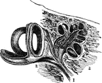
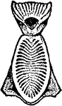
Auditory Nerve
"By anatomists, the auditory nerve is associated with the facial, and is the seventh in order of origin…
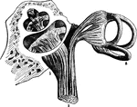
Auditory Nerve
"By anatomists, the auditory nerve is associated with the facial, and is the seventh in order of origin…
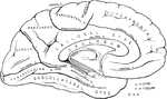
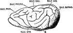
Brain
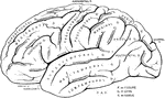
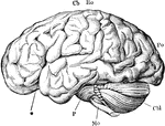
"Profile and vertex views of cerebrum. Dr, the frontal lobe; Par, parietal; Oc, occipital; Ts, temporo-sphenoidal…
Brain
"Diagram illustrating the general relationships of the parts of the brain. A, fore-brain; b, midbrain;…
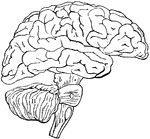
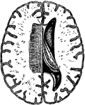
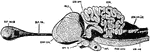
Brain
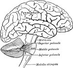
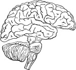
"The brain from the left side. Cb, the cerebral hemispheres forming the main bulkl of the fore-brain;…
Brain
"Diagram of the left half of a vertical median section of the brain. H, H, convoluted inner surface…
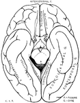
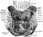
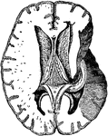
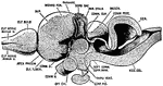
Brain
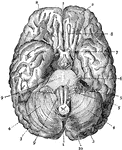
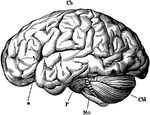
The brain seen from below. 1: Great fissure; 2: Anterior lobes of cerebrum; 3: Posterior lobes of cerebrum;…
Brain
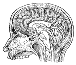
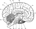
Mesial section through the Corpus Callosum, the Mesencephalon, the Pons, Medulla and Cerebellum. Showing…
Brain
Transverse section through the human mesencephalon at the level of the superior quadrigeminal body


Brain
Orbital surface of the left frontal lobe and the island of Reil; the tip of the temporo-sphenoidal lobe…
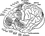
Brain
Horizontal section through the Right Cerebral Hemisphere at the Level of the Widest Part of the Lenticular…
Brain
Diagram illustrating the general relationships of the parts of the brain. Labels: A, fore-brain; b,…
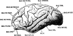
Brain
The brain from the left side. Labels: Cb, the cerebral hemispheres forming the main bulk of the fore-brain;…
Brain
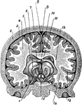
A cross section of the brain from left to right. Labels: 1, thalamus; 2, skull; 3, cerebral membrane;…
Brain and Cranial Nerves
The brain and the cranial nerves seen partly in section and partly in side view. Labels: C, convolutions…
Brain and Spinal Cord
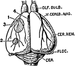
Anterior view of the brain and spinal marrow. Labels: 1, 1, hemispheres of the cerebrum; 2, great middle…
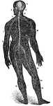
Brain and Spinal Cord
The brain and spinal cord. Labels: 1, 1, hemispheres of cerebrum; 2, great middle fissure; 3, cerebellum;…
Brain and Spinal Cord of Fetus
Human fetus in the third month of development, with the brain and spinal cord exposed from behind.
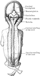
A Back View of the Brain and Spinal Cord
A back view of the brain and spinal cord. Labels: 1, The cerebrum. 2, The cerebellum. 3, The spinal…
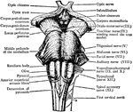
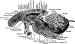
The Brain and the Cranial Nerves
The brain and the origin of the twelve pairs of cranial nerves. Labels: F, E, the cerebrum; D, the cerebellum,…
Fetal Brain at Six Months
Side view of fetal brain at six months, showing commencement of formation of the principal fissures…
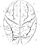
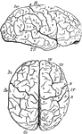
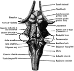
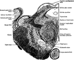
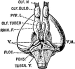
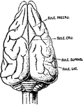
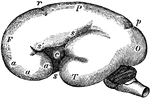
View of Brain from Above
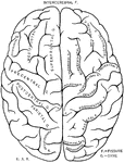
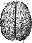
View of brain from above. F, Frontal lobe; P, Parietal lobe; O, Occipital lobe; T, Temporal lobe; S,…
Brain Hemispheres and Spinal Cord
1. Hemispheres of the brain proper, or cerebrum. 2. Hemispheres of the smaller brain, or cerebellum.…
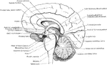
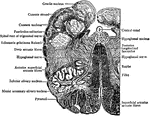
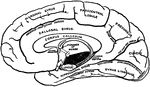
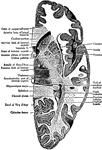
Brain in Mesial Section
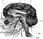
Simplified drawing of brain as seen in mesial section, showing relation of brain stem, cerebrum and…
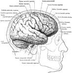
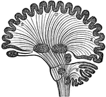
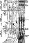
Diagram of Human Brain in Vertical Section
Diagram of human brain in vertical section, showing the situation of the different ganglia and the course…
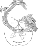
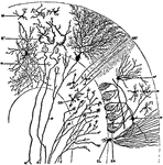
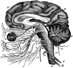
Connection of Brain Nerves
Diagram showing the brain connections of the vagus, glossopharyngeal, auditory, facial, abducent, and…