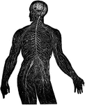
View of the nervous system in man, showing the nervous centers (the brain and the spinal near row) where the other nerves begin which provide the whole body.

Nervous System

View of the nervous system in man, showing the nervous centers (the brain and the spinal near row) where…

"Part of the nervous system viewed in front." —Davison, 1910

Nervous System

"Part of the nervous system viewed in front." —Davison, 1910

"Diagram illustrating the General Arrangement of the Nervous System. (posterior view.)" — Blaisedell, 1904

Nervous System

"Diagram illustrating the General Arrangement of the Nervous System. (posterior view.)" — Blaisedell,…
"A nerve cell from the brain." — Ritchie, 1918

Neuron

"A nerve cell from the brain." — Ritchie, 1918

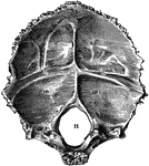
Occipital bone of the human skull, inner surface. It is situated at the back and base of the skull. Labels: 9,9, and 10,10, depression for reception of lobes of brain; 11, foramen magnum.

Occipital Bone of the Human Skull

Occipital bone of the human skull, inner surface. It is situated at the back and base of the skull.…

Diagram of the minute structure of the olfactory bulb.

Minute Structure of Olfactory Bulb

Diagram of the minute structure of the olfactory bulb.

Diagram to illustrate the development of the opercula which cover the insula. A, Sylian fossa before opercula begin to form. B, Fronto-paretal and temporal opercula wells advances. C, All the four opercula developed but not in opposition. Labels: F.P., fronto-parietal operculum; T, temporal operculum; F.R., frontal wall of fossa; O.R., orbital wall of fossa; F., frontal operculum; O, orbital operculum; s2, anterior horizontal limb of Sylvian fissure; s3, ascending limb; s4, posterior horizontal limb.

Development of the Opercula

Diagram to illustrate the development of the opercula which cover the insula. A, Sylian fossa before…

"The size of the hemispheres of the brain (A) is so small that they leave exposed the olfactory ganglion (a), the cerebellum (C), and more or less of the optic lobes (B), and they are but partially connected together by the 'fornix' and 'anterior commissure,' the great cerebral commissure known as the 'corpus callosum' being absent." — Chambers, 1881

Opossum Brain

"The size of the hemispheres of the brain (A) is so small that they leave exposed the olfactory ganglion…
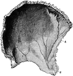
Parietal bone of the human skull, inner surface. The parietal bones form the greater part of the sides and roof of the skull. Labels: A, parietal depression; E, furrow for ramification of arteries.

Parietal Bone of the Human Skull

Parietal bone of the human skull, inner surface. The parietal bones form the greater part of the sides…

"Brain of Perch. Upper aspect. a, cereoellum; b, optic lobes; c, hemispheres; e, lobi inferiores; f, hypophysis; g, lobi posteriores; i, olfactory lobes; n, nervus opticus; o, nervus olfactorius; p, nervus olulo-motorius; g, nervus trochlearis; r, nervus trigeminus; s, nervus acousticus; t, nrvus vagus; u, nervus abducens; v, fourth ventricle." — Encyclopedia Britannica, 1893

Perch Brain

"Brain of Perch. Upper aspect. a, cereoellum; b, optic lobes; c, hemispheres; e, lobi inferiores; f,…

"Brain of Perch. Lower aspect. a, cereoellum; b, optic lobes; c, hemispheres; e, lobi inferiores; f, hypophysis; g, lobi posteriores; i, olfactory lobes; n, nervus opticus; o, nervus olfactorius; p, nervus olulo-motorius; g, nervus trochlearis; r, nervus trigeminus; s, nervus acousticus; t, nrvus vagus; u, nervus abducens; v, fourth ventricle." — Encyclopedia Britannica, 1893

Perch Brain

"Brain of Perch. Lower aspect. a, cereoellum; b, optic lobes; c, hemispheres; e, lobi inferiores; f,…
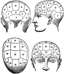
The term applied to the psychological theories of Gall and Spurzheim, founded upon 1, the discovery that the brain, as the organ of the mind, is not so much a single organ as a complex congeries of organs; and 2, observations as to the existence of a certain correspondence between the aptitudes of the individual and the configuration of his skull.

Phrenology

The term applied to the psychological theories of Gall and Spurzheim, founded upon 1, the discovery…
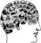
A theory which claims to be able to determine character, personality traits, and criminality on the basis of the shape of the head. This technique was developed in the early 1800's and is currently discredited as a pseudoscience.

Phrenology

A theory which claims to be able to determine character, personality traits, and criminality on the…
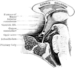
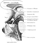
Mesial section through the pituitary region in a child of twelve months old.

Pituitary Region in a Child

Mesial section through the pituitary region in a child of twelve months old.

Mesial section through the pituitary region in an adult.

Pituitary Region in an Adult

Mesial section through the pituitary region in an adult.

"Brain of Polypterus. Upper aspect. a, medulia; b, corpora restiformia; c, cerebellum; d, lobi optici; e, hypophysis; f, fissura cerebri magna; g, nervus opticus; g', chiasma; h, hemispheres; i, lobus olfactorius; k, sinus rhomboidalis (fourth ventricle)." — Encyclopedia Britannica, 1893

Polypterus Brain

"Brain of Polypterus. Upper aspect. a, medulia; b, corpora restiformia; c, cerebellum; d, lobi optici;…

"Brain of Polypterus. Lateral aspect. a, medulia; b, corpora restiformia; c, cerebellum; d, lobi optici; e, hypophysis; f, fissura cerebri magna; g, nervus opticus; g', chiasma; h, hemispheres; i, lobus olfactorius; k, sinus rhomboidalis (fourth ventricle)." — Encyclopedia Britannica, 1893

Polypterus Brain

"Brain of Polypterus. Lateral aspect. a, medulia; b, corpora restiformia; c, cerebellum; d, lobi optici;…

"Brain of Polypterus. Lower aspect. a, medulia; b, corpora restiformia; c, cerebellum; d, lobi optici; e, hypophysis; f, fissura cerebri magna; g, nervus opticus; g', chiasma; h, hemispheres; i, lobus olfactorius; k, sinus rhomboidalis (fourth ventricle)." — Encyclopedia Britannica, 1893

Polypterus Brain

"Brain of Polypterus. Lower aspect. a, medulia; b, corpora restiformia; c, cerebellum; d, lobi optici;…
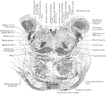
Section through the lower part of the human pons varolii immediately above the medulla.

Section Through Pons Varolii

Section through the lower part of the human pons varolii immediately above the medulla.
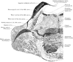
Section through the pons varolii at the level of the nuclei of the trigeminal nerve in an orangoutang.

Section Through Pons Varolii

Section through the pons varolii at the level of the nuclei of the trigeminal nerve in an orangoutang.

Section through the upper part of the pons varolii of the orangoutang above the level of the trigeminal nerve.

Section Through Pons Varolii

Section through the upper part of the pons varolii of the orangoutang above the level of the trigeminal…
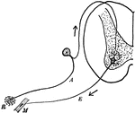
"Diagram of the simple reflex arc. R, receptor; A, afferent (sensory) neuron; E, efferent (motor) meuron; M, muscle." —Martin, 1917

Reflex Arc

"Diagram of the simple reflex arc. R, receptor; A, afferent (sensory) neuron; E, efferent (motor) meuron;…
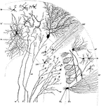
Transverse section through a cerebellar folium. Labels: A, axon of cell Purkinje; F, moss fibers; K and K', fibers from the white core of folium ending in molecular layer in connection with the dendrites of the cells of Purkinje; M, small cell of the molecular layer; GR, granule cell; M1, basket cell; ZK, basket work around the cells of Purkinje; GL, neuroglial cell; N, axon of an association cell.

Sagittal Section Through Cerebellar Folium

Transverse section through a cerebellar folium. Labels: A, axon of cell Purkinje; F, moss fibers; K…
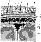
Diagram showing the layers of the scalp and membranes of the brain in section. Labels: a, skin; b, subcutaneous tissue with hair roots and vessels; c, epicranius; d, subepicranial layer; e, pericranium; fm parietal bone; g, dura mater; k, arachnoid; l, pia mater; m, cortex; n, in subdural space near a Pacchionian body projecting within the superior longitudinal sinus.

Layers of the Scalp and Membrane of the Brain

Diagram showing the layers of the scalp and membranes of the brain in section. Labels: a, skin; b, subcutaneous…
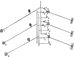
"Diagram to illustrate how a single sensory neuron may communicate with several motor neurons, and a single motor neuron with several sensory neurons." —Martin, 1917

Sensory Neuron

"Diagram to illustrate how a single sensory neuron may communicate with several motor neurons, and a…

Sphenoid bone, situated the anterior part of the base of the skull, articulating with all the other cranial bones, which it binds firmly and solidly together. Labels: a, greater wing; b, lesser wing.

Sphenoid Bone of the Human Skull

Sphenoid bone, situated the anterior part of the base of the skull, articulating with all the other…

Two sections through the tegmentum of the pons at its upper part, close to the mesencephalon. A is at a slightly lower level than B.

Section Through Tegmentum

Two sections through the tegmentum of the pons at its upper part, close to the mesencephalon. A is at…
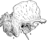
Temporal bone of the human skull. The temporal bones are situated at the sides and base of the skull. Labels: 1, squamous portion; 2, placed below external opening of auditory canal in petrous portion; 3, placed below mastoid portion; 4, placed below glenoid cavity for reception of condyle of lower jaw.

Temporal Bone of the Human Skull

Temporal bone of the human skull. The temporal bones are situated at the sides and base of the skull.…
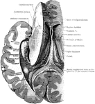
Dissection, to show the fornix and the posterior and descending cornua of the lateral ventricle of the left side.

Dissection to Show Ventricle and Fornix

Dissection, to show the fornix and the posterior and descending cornua of the lateral ventricle of the…
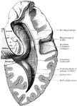
Dissection, to show the posterior and descending cornua of the lateral ventricle. Labels: B.G., Giacomini's band; F.D., gyrus dentatus; F, fimbria; H.C., hippocampal convolution.

Dissection to Show Ventricle

Dissection, to show the posterior and descending cornua of the lateral ventricle. Labels: B.G., Giacomini's…
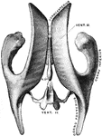
Drawing taken from a cast of the ventricular system of the brain, as seen from above. Vent. III, Third ventricle. Vent. IV, Fourth Ventricle. R.SP., recessus suprapinealis.

Ventricles of the Brain

Drawing taken from a cast of the ventricular system of the brain, as seen from above. Vent. III, Third…
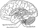
Scheme showing relations of the ventricles to the surface of the brain.

Relations of the Ventricles to the Surface of the Brain

Scheme showing relations of the ventricles to the surface of the brain.
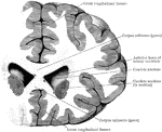
Coronal section through the frontal lobes and the anterior horns of the lateral ventricles.

Section Through Lateral Ventricles

Coronal section through the frontal lobes and the anterior horns of the lateral ventricles.