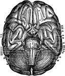
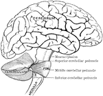
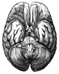
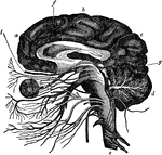
Base of the brain and cerebellum, together with the cranial nerves.

Base of Brain and Cerebellum

Base of the brain and cerebellum, together with the cranial nerves.

Part of the central nervous system found in the skull.

Brain

Part of the central nervous system found in the skull.

Scheme showing the connection of the several parts to the brain.

Brain

Scheme showing the connection of the several parts to the brain.

"Diagram illustrating the general relationships of the parts of the brain. A, fore-brain; b, midbrain; B, cerebellum; C, pons Varolii; D, medulla oblongata; B, C, and D together constitute the hind-brain." —Martin, 1917

Brain

"Diagram illustrating the general relationships of the parts of the brain. A, fore-brain; b, midbrain;…
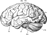
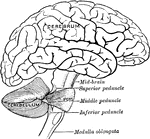
"The brain from the left side. Cb, the cerebral hemispheres forming the main bulkl of the fore-brain; Cbl, the cerebellum; Mo, the medulla oblongata; P, the pons Varolii; *, the fissure of Sylvius; Ro, the fissure of Rolando; Po, the Parieto-occipital fissure." —Martin, 1917

Brain

"The brain from the left side. Cb, the cerebral hemispheres forming the main bulkl of the fore-brain;…
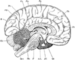
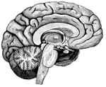
"Diagram of the left half of a vertical median section of the brain. H, H, convoluted inner surface of left cerebral hemisphere; Cc, corpus callosum; Th, optic thalamus; c.q., corpora quadrigemina; Cb, cerebellum; Sp.c, spinal cord; Mo, medulla oblongata; P, pons Varolii; oc, oculo-motor nerve; pt, pituitary body; op, optic nerve; Ro, fissure of Rolando; Po, parieto-occipital fissure; Fr, frontal lobe; Pa, parietal lobe; O, occipital lobe." —Martin, 1917

Brain

"Diagram of the left half of a vertical median section of the brain. H, H, convoluted inner surface…
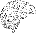
Diagram illustrating the general relationships of the parts of the brain. Labels: A, fore-brain; b, mid-brain; B, cerebellum; C, pons Varolii; D, medulla oblongata; B,c,and D together constitute the hind-brain.

Brain

Diagram illustrating the general relationships of the parts of the brain. Labels: A, fore-brain; b,…
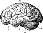
The brain from the left side. Labels: Cb, the cerebral hemispheres forming the main bulk of the fore-brain; Cbl, the cerebellum; Mo, the medulla oblongata; P, the pons Varolii; *, the fissure of Sylvius.

Brain

The brain from the left side. Labels: Cb, the cerebral hemispheres forming the main bulk of the fore-brain;…

Anterior view of the brain and spinal marrow. Labels: 1, 1, hemispheres of the cerebrum; 2, great middle fissure; 3, cerebellum; 4, olfactory nerves; 5, optic nerves; 6, corpora albicantia; 7, motor oculi nerves; 8, pons Varolii; 9, fourth pair of nerves; 10, lower portion of the medulla oblongata; 11, 11, medulla spinalis in its whole length; 12, 1, spinal nerves; 13, cauda equina.

Brain and Spinal Cord

Anterior view of the brain and spinal marrow. Labels: 1, 1, hemispheres of the cerebrum; 2, great middle…

Human fetus in the third month of development, with the brain and spinal cord exposed from behind.
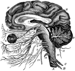
Brain and Spinal Cord of Fetus

Human fetus in the third month of development, with the brain and spinal cord exposed from behind.

The brain and the origin of the twelve pairs of cranial nerves. Labels: F, E, the cerebrum; D, the cerebellum, showing the arbor vitae; G, the eye; H, the medulla oblongata; A, the spinal cord; C and B, the first two pairs of spinal nerves.

The Brain and the Cranial Nerves

The brain and the origin of the twelve pairs of cranial nerves. Labels: F, E, the cerebrum; D, the cerebellum,…

Simplified drawing of brain as seen in mesial section, showing relation of brain stem, cerebrum and cerebellum, and ventricular spaces.

Brain in Mesial Section

Simplified drawing of brain as seen in mesial section, showing relation of brain stem, cerebrum and…
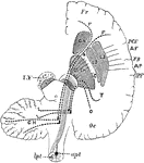
Schema showing the connections of the several parts of the brain.

Connection of the Brain Parts

Schema showing the connections of the several parts of the brain.

Diagram to show the connecting of the Frontal Occipital Lobes with the Cerebellum. The dotted lines passing in the crusta, outside the motor fibers, indicate the connection between the temporo-occipital lobe and the cerebellum. F.C., the fronto-cerebellar fibers, which pass internally to the motor tract in the crusta; I.F., fibers from the caudate nucleus to the pons. FR., frontal lobe; Oc, occipital lobe; AF. ascending frontal; AP., ascending frontal convolution; FR., fissure of Rolando; IPF., interparietal fissure, a section of crus is lettered on the left side. SN., substantia nigra; PY. pyramidal motor fiber, which on the right is shown as continuous lines converging to pass through the posterior limb of IC. internal capsule ( the knee or elbow of which is shown thus) upwards into the hemisphere and downwards through the pons to cross at the medulla in the anterior pyramids.

Brain Showing Connection of Frontal Occipital Lobe with Cerebellum

Diagram to show the connecting of the Frontal Occipital Lobes with the Cerebellum. The dotted lines…
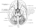
Simplified drawing of brain as seen from below, showing relations of brain stem to spinal cord and cerebrum.
Relation of Brain Stem to Spinal Cord

Simplified drawing of brain as seen from below, showing relations of brain stem to spinal cord and cerebrum.

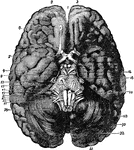
The base of the brain. Labels: 1, longitudinal fissure; 2, 2, anterior lobes of cerebrum; 3, olfactory bulb; 7, optic commissure; 9, 3rd nerve, ; 11, 4th nerve; 13, 5th nerve; 14, crura cerebri, 15, 6th nerve; 16, pons Varolii; 17, 7th nerve; 19, 8th nerve; 20, medulla oblongata; 21, 9th nerve, 23, 10th nerve; 25, 11th nerve; 27, 12th nerve; 28, 29, 30, 31, 32, cerebellum.

Base of Brain

The base of the brain. Labels: 1, longitudinal fissure; 2, 2, anterior lobes of cerebrum; 3, olfactory…
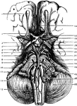
The base of brain. Labels: 1. Olfactory Bulb; 2. Second, or Optic Nerves; 3. Anterior Perforated Space; 4. Optic Tract; 5. Crus Cerebri; 6. 3rd Nerve; 7. 4th Nerve.; 8. 5th Nerve; 9. 6th Nerve; 10. Pyramid; 11. Olivary Body; 12. Vertebral Artery; 13. Anterior Spinal Artery; 14. Anterior Cerebral Artery; 15. Lamina Cinerea; 16. Middle Cerebral Artery; 17. Tuber Cinereum; 18. Corpora Albicantia; 19. Posterior Perforated Space; 20. Posterior Cerebral Artery; 21. Superior Cerebral Artery; 22. Pons Varolii; 23. Inferior Cerebellar Artery; 24. 7th and 8th Nerves; 25. 9th, 10th, and 11th Nerves; 26. 12th Nerve; 27. Cerebellum.

Base of the Brain

The base of brain. Labels: 1. Olfactory Bulb; 2. Second, or Optic Nerves; 3. Anterior Perforated Space;…
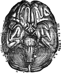
Base of the Brain. Labels: 1,2, longitudinal fissure; 3, anterior lobes cerebrum; 4, middle lobe; 5, fissure Sylvius; 6, posterior lobe; 7, infundibulum; 8, its body; 9, corporo albicantia; 10, cineritious matter; 11, crura cerebri; 12, pons Varolii; 13, medulla oblongata; 14, posterior prolongation of pons; 15, middle of cerebellum; 16, anterior part of cerebellum; 17, its posterior part and fissure; 18, medulla spinalis; 19, middle fissure medulla; 20, pyramidal body; 21, retiform body; 22, olivary body; 23, olfactory nerve; 24, its bulb; 25, its external root; 26, middle root; 27, internal root; 28, 29, optic nerve; 30, third nerve; 31, fourth nerve; 32, fifth nerve; 33, sixth nerve; 34, facial nerve; 35, auditory nerve; 36, 37, 38, eighth nerve.

Base of the Brain

Base of the Brain. Labels: 1,2, longitudinal fissure; 3, anterior lobes cerebrum; 4, middle lobe; 5,…

Vertical section of dog's cerebellum. Labels: p m, pia mater; p, corpuscles of Purkinje, which are branched nerve cells lying in a single layer and sending single processed downwards and more numerous ones upwards, which branch continuously and extend through the deep "molecular layer" towards the free surface; g, dense layer of ganglionic corpuscles., closely resembling nuclear layers of retina; f, layer of nerve fibers, with a few scattered ganglionic corpuscles. This last layer (f) constitutes part of the white matter of the cerebellum, while the layer between it and the free surface are gray matter.

Cerebellum of Dog's Brain

Vertical section of dog's cerebellum. Labels: p m, pia mater; p, corpuscles of Purkinje, which are branched…

Outline sketch of a section of the cerebellum, showing the corpus dentatum. The section has been carried through the left lateral part of the pons, so as to divide the superior peduncle and pass nearly through the middle of the left cerebellar hemisphere. The olivary body has also been divided longitudinally so as to expose in section its corpus dentatum. c r, crus cerebri; f, fillet; q, corpora quadrigemina; s p, superior peduncle of the cerebellum divided; m p, middle peduncle or lateral part of the pons Varolii, with fibers passing from it into the white stem; a v, continuation of the white stem radiating towards the arbor vitae of the folia; c d, corpus dentatum; o, olivary body with its corpus dentatum; p, anterior pyramid.

The Cerebellum of the Brain

Outline sketch of a section of the cerebellum, showing the corpus dentatum. The section has been carried…
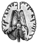
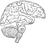
Top view of the human brain.

Human Brain

Top view of the human brain.

A top view of a dissection of the human brain showing the lateral fourth and fifth ventricles.

Human Brain

A top view of a dissection of the human brain showing the lateral fourth and fifth ventricles.

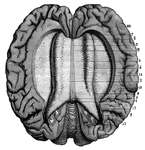
A view of the Corpus Callosum from above.

Human Brain

A view of the Corpus Callosum from above.

Side diagram of the human brain showing which parts of the brain control hearing, speech, vision, legs, head, arm, and face.

Human Brain

Side diagram of the human brain showing which parts of the brain control hearing, speech, vision, legs,…

Side diagram of the human brain showing which areas perform the sense of taste, smell, and vision.
Human Brain

Side diagram of the human brain showing which areas perform the sense of taste, smell, and vision.

"<em>A</em>,frontal love of the cerebrum; <em>B</em>, parietal lobe; <em>C</em>, parieto-occipital lobe; <em>D</em>, occcipital lobe; <em>E</em>, cerebellum; <em>F</em>; arbor vitae; <em>H</em>, pons Varolii; <em>K</em>, medulla oblongata." — Blaisedell, 1904

Left Half of the Brain

"A,frontal love of the cerebrum; B, parietal lobe; C, parieto-occipital lobe;…
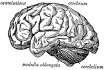
"The brain seen from the side, showing the three principal divisions." &mdash; Ritchie, 1918

Side view of the brain

"The brain seen from the side, showing the three principal divisions." — Ritchie, 1918
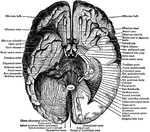
The general arrangement of the brain and attached nerves and blood vessels. At <em>a b</em> and <em>c</em> is the cerebrum; at <em>f</em> is represented the corpus callosum that joins the two hemispheres together; at d is the cerebellum; at <em>g</em> is the beginning of the optic nerves and at <em>l</em> is the olfactory nerve.

The Brain

The general arrangement of the brain and attached nerves and blood vessels. At a b and c

View of the under surface of the brain, with the lower portion of the temporal and occipital lobes, and the cerebellum on the left side removed, to show the origins of the cranial nerves.

Under Surface of the Brain

View of the under surface of the brain, with the lower portion of the temporal and occipital lobes,…
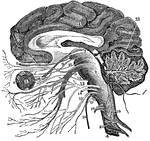
A vertical section of the cerebrum, cerebellum, and the medulla oblongata, showing the relation of the cranial nerves at their origin. Labels: 1, The cerebrum. 2, The cerebellum with its arbor vitae represented. 3, The medulla oblongata. 4, The spinal cord. 5, The corpus callosum. 6, The first pair of nerves. 7, The second pair. 8, The eye. 9, The third pair of nerves. 10, The fourth pair. 11, The fifth pair. 12, The sixth pair. 13, The seventh pair. 14, The eighth pair. 15, The ninth pair. 16, The tenth pair. 17, The eleventh pair. 18, The twelfth pair. 19, Spinal nerves. 21, The tentorium.

Vertical Section of the Brain

A vertical section of the cerebrum, cerebellum, and the medulla oblongata, showing the relation of the…

"Brain of Carcharias. ae, nervus acousticus; b, corpus restiforme; c, cerebellum; d, lobus opticus; e, hypophysis; g, nervus opticus; h, hemisphere; i, lobus olfactorius; i', olfactory pedicle; k, nervus olfactorius; l, epiphysis; m, nervus oculo-motorius; tr, nervus trigeminus; v, nervus vagus." &mdash; Encyclopedia Britannica, 1893

Carcharias Brain

"Brain of Carcharias. ae, nervus acousticus; b, corpus restiforme; c, cerebellum; d, lobus opticus;…
"Diagram showing the position of the nervous centers in the head." &mdash; Tracy, 1888

Cerebellum

"Diagram showing the position of the nervous centers in the head." — Tracy, 1888

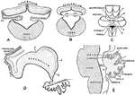
Showing the development of the cerebellum. A, Transverse section through the forepart of the cerebellum of a sheep embryo. B, Transverse section through the hinder part of the cerebellum of a sheep embryo. C, Cerebellum of a human fetus 17 cm long. D, Median section through cerebellum of sheep embryo 5 cm long.E, Median section through cerebellum of human fetus 17 cm long. Labels: 1, sulcus primarius; 2, sulcus above and afterwards behind the pyramid; 3, sulcus between pyramid and uvula; 4, great horizontal fissure; r.l., lateral recess ventricle IV; T, transverse groove in roof of ventricle IV.

Development of Cerebellum

Showing the development of the cerebellum. A, Transverse section through the forepart of the cerebellum…

The lower surface of the cerebellum. The tonsil on the right side has been removed so at to display more fully the inferior medullary velum and the furrowed band.

Lower Surface of Cerebellum

The lower surface of the cerebellum. The tonsil on the right side has been removed so at to display…

Sagittal section through the left lateral hemisphere of the cerebellum. Showing the "arbor vitae" and the corpus dentatum.

Sagittal Section Through Cerebellum

Sagittal section through the left lateral hemisphere of the cerebellum. Showing the "arbor vitae" and…
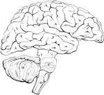
The upper surface of the cerebellum.

Upper Surface of Cerebellum

The upper surface of the cerebellum.

Plan in outline of the encephalon, as seen from the right side. The parts are represented as separated from one another somewhat more than natural, so as to show their connections. A, cerebrum; f, g, h, its anterior middle, and posterior lobes; e, fissure of Sylvius; B, cerebellum; C, pons Varolii; D, medulla oblongata; a, peduncles of the cerebellum; C, pons Varolii; D, medulla oblongata; A, peduncles of the cerebrum; b, c, d, superior, middle, and inferior peduncles of the cerebellum.

Outline of the Encephalon

Plan in outline of the encephalon, as seen from the right side. The parts are represented as separated…
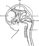
"Diagram of head and brain of human foetus six weeks old (heavy boundaries). The dotted line indicates the outline of the brain of a foetus three months old. Note thee great growth of the hemisphere (h). cer, cerebellum; med, medulla oblongata; mes, mesencephalon; p, pituitary body; pr, prosencephalon; s.c., spinal cord; th, thalamencephalon; 1, olfactory nerve; 2, optic nerve." -Galloway, 1915

Human Fetus

"Diagram of head and brain of human foetus six weeks old (heavy boundaries). The dotted line indicates…
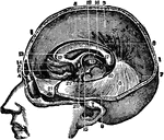
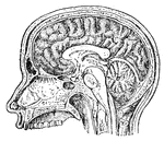
Section of the head showing the greater scythe, the horizontal apophysis of the dura mater between the brain and the cerebellum and other parts under the middle line of the head.

Head, Section of

Section of the head showing the greater scythe, the horizontal apophysis of the dura mater between the…
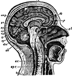
"The Brain is the encephalon, or center of the nervous system and the seat of consciousness and volition in man and the higher animals. Median Longitudinal Section through Head and Upper Part of Neck, to Show relation of Brain to Cranium and the Spinal Cord. <em>c,</em> cerebrum; <em>cb,</em> cerebellum; <em>sc,</em> spinal cord; <em>spc,</em> spinal column; <em>mo,</em> medulla oblongata passing, through foramen magnum, into the spinal cord; <em>pv,</em> pons Varolii; <em>cp,</em> cerebral peduncles or crura cerebri; <em>cqa,</em> anterior corpora quadrigemina; <em>cqp,</em> posterior corpora quadrigemina; <em>pg,</em> pineal gland; <em>pb,</em> pituitary body; <em>cc,</em> corpus collosum, divided transversely; <em>f,</em> fornix; <em>mg,</em> marginal gyrus; <em>gf,</em> gyrus fornicatus; <em>cmg,</em> calloso-marginal suleus; <em>O,</em> occipital lobe; <em>po,</em> parieto-occipital fissure; <em>cf,</em> calcarine fissure; <em>dm,</em>dura mater, separating cerebrum from cerebellum."&mdash;(Charles Leonard-Stuart, 1911)

Human Brain

"The Brain is the encephalon, or center of the nervous system and the seat of consciousness and volition…

"Part of the nervous system viewed in front." —Davison, 1910

Nervous System

"Part of the nervous system viewed in front." —Davison, 1910

"The size of the hemispheres of the brain (A) is so small that they leave exposed the olfactory ganglion (a), the cerebellum (C), and more or less of the optic lobes (B), and they are but partially connected together by the 'fornix' and 'anterior commissure,' the great cerebral commissure known as the 'corpus callosum' being absent." &mdash; Chambers, 1881

Opossum Brain

"The size of the hemispheres of the brain (A) is so small that they leave exposed the olfactory ganglion…

"Brain of Polypterus. Upper aspect. a, medulia; b, corpora restiformia; c, cerebellum; d, lobi optici; e, hypophysis; f, fissura cerebri magna; g, nervus opticus; g', chiasma; h, hemispheres; i, lobus olfactorius; k, sinus rhomboidalis (fourth ventricle)." &mdash; Encyclopedia Britannica, 1893

Polypterus Brain

"Brain of Polypterus. Upper aspect. a, medulia; b, corpora restiformia; c, cerebellum; d, lobi optici;…

"Brain of Polypterus. Lateral aspect. a, medulia; b, corpora restiformia; c, cerebellum; d, lobi optici; e, hypophysis; f, fissura cerebri magna; g, nervus opticus; g', chiasma; h, hemispheres; i, lobus olfactorius; k, sinus rhomboidalis (fourth ventricle)." &mdash; Encyclopedia Britannica, 1893

Polypterus Brain

"Brain of Polypterus. Lateral aspect. a, medulia; b, corpora restiformia; c, cerebellum; d, lobi optici;…

"Brain of Polypterus. Lower aspect. a, medulia; b, corpora restiformia; c, cerebellum; d, lobi optici; e, hypophysis; f, fissura cerebri magna; g, nervus opticus; g', chiasma; h, hemispheres; i, lobus olfactorius; k, sinus rhomboidalis (fourth ventricle)." &mdash; Encyclopedia Britannica, 1893

Polypterus Brain

"Brain of Polypterus. Lower aspect. a, medulia; b, corpora restiformia; c, cerebellum; d, lobi optici;…

"Partial section of a Vertebrate brain (diagrammatic). OLF., Olfactory lobe; CH., cerebral hemispheres; C., wall of cerebrum cut to show ventricle, behind this the figure is that of a median section; PA., parietal organ arising from thalamencephalon; Pl., pineal organ; INF., infundibulum descending from thalamencephalon; H., hypophysis; OL., optic lobes; CB., cerebellum; CPL., choroid plexus on roof of fourth ventricle; MO., floor of the medulla oblongata; CC., central canal of spinal cord." -Thomson, 1916

Vertebrate Brain

"Partial section of a Vertebrate brain (diagrammatic). OLF., Olfactory lobe; CH., cerebral hemispheres;…