Pharynx and EsophagusClipart ETC: An online service of Florida's Educational Technology Clearinghouse

Pharynx and Esophagus

Pharynx and EsophagusPharynx and EsophagusPharynx and Esophagus

To use any of the clipart images above (including the thumbnail image in the top left corner), just click and drag the picture to your desktop. You may also control-click (Mac) or right-click (Windows) and choose "Save file to disk" from the pop-up menu. Click here for help downloading and using clipart files.

Download TIFF* File

* A TIFF file is a high-resolution (240 dpi) format. TIFF files should be used only if you are printing the clipart. Do NOT use TIFF files for clipart that will displayed on a computer screen. Click here for help downloading and using TIFF files.

File Name: 56716_esophagus
Description: 
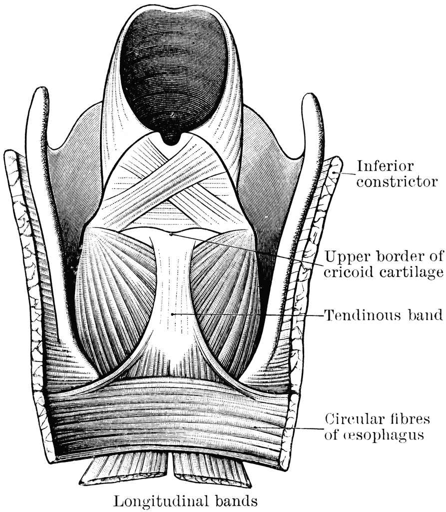
The lower part of the pharynx and the upper part of the esophagus have been slit up from behind, and the mucous membrane removed to show the muscular fibers. The two longitudinal bands are seen coming round to the front to be attached by a common tendon to the upper border of the cricoid cartilage.
Source: Cunningham, D.J. Textbook of Anatomy (New York: William Wood and Co., 1903) 992
Keywords: esophagus, stomach, hyoidm trachea, cricoid, thyroid

Copyright: 2009, Florida Center for Instructional Technology. See license.

Main MenuSite MapSearchLicenseClipart Help


Clipart ETC is a part of the Educational Technology Clearinghouse and is funded by various grants. Produced by the Florida Center for Instructional Technology, College of Education, University of South Florida. Email the project manager.


Clipart ETCEducational Technology Clearinghouse