Abdomen
"The belly; that part of the body of a mammal which lies between the thorax and the pelvis; In entomology,…
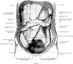
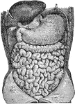
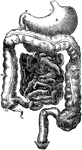
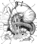
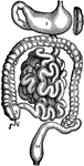
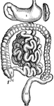
Abdomen Laid Open After Removal of Jejunum and Ileum
The abdomen viscera after the removal of the jejunum and ileum. The transverse colon is much more regular…
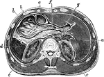
Horizontal Section Through Abdomen
Horizontal section through upper part of abdomen. Labels: a, liver; b, stomach; c, transverse colon;…
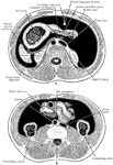
Transverse Section of Abdomen
Diagrammatic transverse section of abdomen, to show the peritoneum on transverse tracing. A, at level…
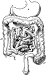
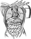
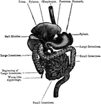
Abdominal Organs
Abdominal organs. Labels: 1, liver turned up; 2, gall bladder; 3, stomach; 4, large intestine; 5, small…
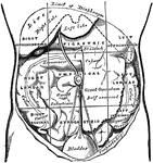
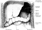
Regions of the Abdomen and their Contents
Regions of the abdomen and their contents (edge of costal cartilages in dotted outline). "For convenience…
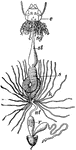
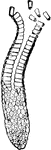
Ailmentary Canal
"ailmentary canal of a honey bee. at, honey stomach; s, true stomach; nt, intestine; o, esophagus; sg,…
Alimentary Canal
"The digestive sac, tract, or tube of any animal; the visceral or intestinal cavity."-Whitney, 1902
Alimentary Canal
Diagram of the abdominal part of the alimentary canal (digestive system). Labels: C, the cardiac, and…
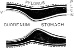
Alimentary Canal
Diagram of the abdominal part of the alimentary canal. Labels: C, the cardiac, and P, the pyloric end…
General View of the Alimentary Canal
Labels: O, esophagus; S, stomach; SI, small intestine; LI, large intestine, Sp spleen; L, liver (raised…
Anemone
"Sea anemone dissected; c, tentacles; d, mouth; e, stomach; white lines above k, the mesenteries." —Davison,…
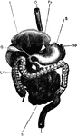
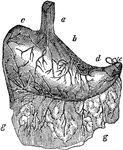
Arteries of the Abdominal Organs
Arteries of the abdominal organs. Labels: 1, the liver; 2, the stomach; 3, upper gut; 4, pancreas; 6,…

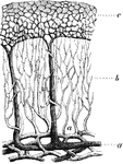
Blood Vessels in the Mucous Membrane of the Stomach
Termination of the blood vessels in the mucous membrane of the stomach.
Blood Vessels of the Stomach
Plan of the blood vessels of the stomach, as they would be seen in a vertical section. Labels: a, arteries,…
Columnar Epithelium
"In the stomach, intestines and elsewhere the epithelial cell is obling in profile and is called columnar…
Crop and Digestive Organs
The swelling under the throat is called the crop, or first stomach. It is largely developed in some…
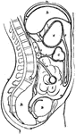
Cunina Rhododactyla
"Diagram of a vertical section through a young Cunina rhododactyla, passing on the right side through…
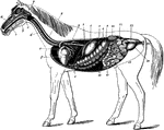
Digestive Apparatus of the Horse
The digestive apparatus of the horse. Labels: a, mouth; 2, pharynx; 3, esophagus; 4, diaphragm; 5, spleen;…
Digestive Organs
The stomach, pancreas, liver, and duodenum, with part of the rest of the small intestine and the mesentery;…
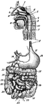
Digestive Organs of a Dog
Stomach, liver, pancreas, and duodenum of a dog. Labels: a, liver; b, gall bladder; c, biliary canals;…
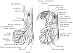
Digestive Organs of a Horse
The relation of anterior abdominal digestive organs- left antero-lateral view. Labels: 1, liver; 2,…
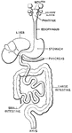
Digestive System
The stomach and intestines. Labels: 1, stomach; 5, 7, 8, 9, 10, 11, large intestine; 3, small intestine;…
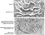
Gastric gland
"The inner coat of the stomach has its surface honeycombed with millions of little pits. We have all…
Gastric Gland from Fundus
Deeper portion of gastric glands from fundus, showing two varieties of lining cells and secretion capillaries…
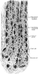
Section through the Gastric Mucous Membrane
A thin section through the gastric mucous membrane which lines the stomach, perpendicular to its surface,…

Girl Laying on Stomach & Reading a Book
An illustration of a girl laying on her stomach and reading a book.
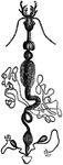
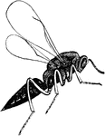
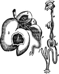
Digestive Apparatus of an Insect
"a, head, antennae, &c; b, pharynx; c, crop; d, gizzard; e, chyle-forming stomach; f, biliary vessels;…
Development of the Intestinal Canal
Two diagrams to illustrate the development of the intestinal canal. The figure to the right shows the…
Mucous Membrane
A portion of the stomach, showing its internal surface or mucous coat. Mucous membranes line various…
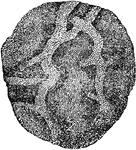
Mucous Membrane of the Stomach
The mucous membrane of the stomach. The top image is natural size and shows the rugae and he mamillated…
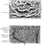
Stomach Mucous Membrane
The mucous membrane of the stomach. A, Natural size. B. Magnified. In A the rugae and the mammilated…
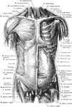
Anterior View of the Muscles of the Trunk
Superficial and deep muscles of the trunk. The sternocleidomastoid, pectoralis major, anterior portion…
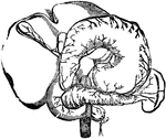
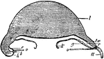
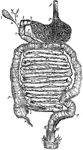
Ox Stomach
"Compound stomach of ox. a, esophagus; b, rumen, or paunch; c, reticulum, or second stomach; d, omasum,…
The Peritoneum
Diagram of the peritoneum, a serous membrane covering all the contents of the abdominal cavity. Labels:…
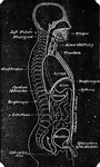
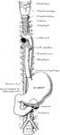
Pharynx and Esophagus
The lower part of the pharynx and the upper part of the esophagus have been slit up from behind, and…
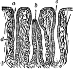
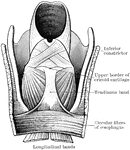
Formation of Pylorus
Diagram to show formation of pylorus. Labels: P, peritoneum; L, longitudinal layer of muscular fibers;…
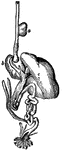
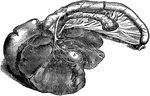
Shark Stomach
"Siphonal stomach and spiral valve of Basking-shark (Selache). a, esophagus; b, cardiac portion of stomach;…
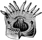
Sheep, Stomachs of
The four stomachs of the sheep, a grass-eating animal. The beginning of the intestines are also shown,…
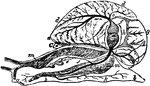
Snail Anatomy
"Anatomy of the Snail: a, the mouth; bb, foot; c, anus; dd, lung; e, stomach, covered above by the salivary…

Stomach
"The stomach is a half-gallon sac, with an outer wall of muscle lined within by mucous membrane, made…
Stomach
The stomach and intestines. 1: Stomach. 2: Duodenum. 3: Small intestine. 4: Termination of the ileum.…
Stomach
Section of the Stomach. 1: Cardiac orifice; 2: Folds of mucous membrane; 3: Muscular wall; 4: Pylorus;…
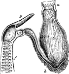
The Stomach
The stomach. Labels: d, lower end of the gullet; a, position of the cardiac aperture; b, the fundus;…
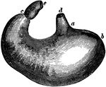
Stomach
The human stomach. Labels: a, the esophagus or gullet; b, the cardiac portion; c, the left extremity;…
Stomach
The digestive system. This figure represents the whole tract of the intestinal canal, not exactly in…
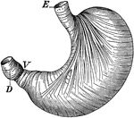
Stomach
The stomach showing the muscles which churn the food. Labels: E, where food enters; V, entrance into…

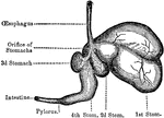
The Stomach and Intestines
The stomach and intestines. Labels: 1, stomach; 2, duodenum; 3, small intestine; 4, termination of the…
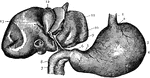
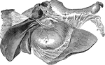
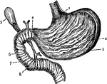
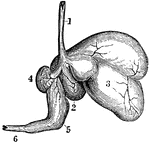
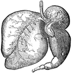
Stomach and Liver
The Stomach and Liver. 1: Esophagus; 2: Cardiac entrance; 3: Large end of stomach; 4: Central portion;…
Stomach Muscles
The three layers of the muscular coat of the stomach. A, Outer or longitudinal layer. B, Middle or circular…
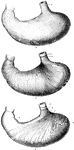
The Stomach of a Sheep
Ruminants (those animals that chew the cud), as the sheep, have a stomach with four cavities. Labels:…
Stomach of an Ox
Ruminants (those animals that chew the cud), as the ox, have a stomach with four cavities. Labels: 1,…