"It consists of a common metallic syringe, A, screwed to a cylindrical valve box, B, which contains two ovoid cavities, in each of which there is a loose, spherical metallic valve. The ends of the cavities are peirced, and the valves fit exactly, either of the orifices. Thus it makes no difference which end of the valve-box is upturned, the valve falling down and closing the orifices in either direction. The flexible india rubber tubes, C D, are attached to the opposite ends of the cavities." —Comstock, 1850

Stomach Pump

"It consists of a common metallic syringe, A, screwed to a cylindrical valve box, B, which contains…

Stomach turned inside out, showing dissection of oblique and circular muscular coats.

Stomach Turned Inside Out

Stomach turned inside out, showing dissection of oblique and circular muscular coats.
Wall of human stomach. Labels: E, epithelium; G, glands; Mm, muscularis mucosae.

Stomach Wall

Wall of human stomach. Labels: E, epithelium; G, glands; Mm, muscularis mucosae.

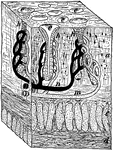
"A tiny block out of the stomach wall. a, the mucous membrane; c and d, the muscles; h, gastric glands; m and n, blood tubes to the glands; e, mouths of glands within the stomach." —Davison, 1910

Cross-Section of Stomach Wall

"A tiny block out of the stomach wall. a, the mucous membrane; c and d, the muscles; h, gastric glands;…
Section through wall of stomach, cardiac portion.

Section Through Stomach Wall

Section through wall of stomach, cardiac portion.

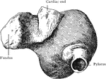
Stomach with puckered fundus, seen from behind and somewhat from left; hardened by formalin.

Stomach with Puckered Fundus

Stomach with puckered fundus, seen from behind and somewhat from left; hardened by formalin.
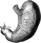
The middle and deep muscular layer of the stomach, viewed from above and in front.

Deep Muscles of the Stomach

The middle and deep muscular layer of the stomach, viewed from above and in front.

Moderately distended stomach, viewed A, from in front; B, from inner or right side; and C, from the outer or left side. The contents of the stomach were carefully removed, through an artificial opening , and replaced with gelatin, the stomach remaining in situ throughout the operation. After the jelly hardened, its exact orientation was carefully noted, and pins indicating the vertical, horizontal, and transverse planes, having been inserted, the organ was removed.

Distended Stomach

Moderately distended stomach, viewed A, from in front; B, from inner or right side; and C, from the…
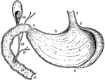
Vertical and longitudinal section of stomach, gall-bladder, and duodenum. Labels: 1, esophagus; 2, cardiac orifice of stomach; 5, lesser curvature; , greater curvature; 8, rugae in interior of stomach; 9, pyloric orifice; 10, 11, 13, interior of duodenum, showing valvulae conniventes; 12, duct conveying bile, and P, duct conveying pancreatic juice, into the duodenum; 14, gall-bladder; 15, commencement of jejunum.

Vertical and Longitudinal Section of Stomach, Gall-Bladder, and Duodenum

Vertical and longitudinal section of stomach, gall-bladder, and duodenum. Labels: 1, esophagus; 2, cardiac…
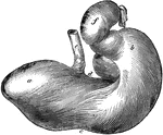
Posterior view of the stomach of a horse. Labels: a, left cul-de-sac; b, right cul-de-sac; c, greater curvature; d, lesser curvature; e, esophagus; f, duodenum.

Horse Stomach

Posterior view of the stomach of a horse. Labels: a, left cul-de-sac; b, right cul-de-sac; c, greater…
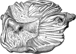
Internal aspect of a horse stomach, opened from below. Labels: a, cuticular mucous membrane; b, villous mucous membrane; c, line of demarcation between the two portions; d, cardiac orifice; e, pyloric orifice and valve.

Horse Stomach

Internal aspect of a horse stomach, opened from below. Labels: a, cuticular mucous membrane; b, villous…
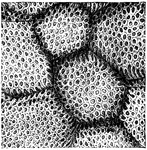
"The Inner Surface of the Stomach, from which the the Epithelium has been removed, showing the Openings of Gastric Glands." — Blaisedell, 1904

Inner surface of the stomach

"The Inner Surface of the Stomach, from which the the Epithelium has been removed, showing the Openings…
The interior of the stomach.

Interior of the Stomach

The interior of the stomach.

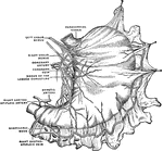
General view of the subperitoneal lymphatic plexus of the stomach prepared by the method of Gerota.
Lymphatic Plexus of the Stomach

General view of the subperitoneal lymphatic plexus of the stomach prepared by the method of Gerota.

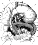
The stomach, pancreas, liver, and duodenum, with part of the rest of the small intestine and the mesentery; the stomach and liver have been turned up so as to expose the pancreas. Labels: V, stomach; D, D',D'', duodenum; L, spleen; P, pancreas; R, right kidney; T, jejunum; Vf, gall bladder; h, hepatic duct; c, cystic duct; ch, common bile duct; 1, aorta; 2, an artery (left coronary) of the stomach; 3, hepatic artery; 4, splenic artery; 5, superior mesenteric artery; 6, superior mesenteric vein; 7, splenic vein; Vp, portal vein.

The Stomach, Pancreas, Liver, and Duodenum

The stomach, pancreas, liver, and duodenum, with part of the rest of the small intestine and the mesentery;…
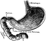
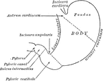
Outlines of the stomach, showing its anatomical landmarks.

Parts of the Stomach

Outlines of the stomach, showing its anatomical landmarks.

The opening part of the stomach where the esophagus joins it is called the cardiac opening; the one at the opposite end, where the small intestine joins it, is called the pylorus.

A Section of the Stomach

The opening part of the stomach where the esophagus joins it is called the cardiac opening; the one…
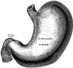
The superficial muscular layer of the stomach, viewed from above and in front.

Superficial Muscles of the Stomach

The superficial muscular layer of the stomach, viewed from above and in front.
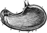
The stomach, the principal organ of digestion. It is a dilated part of the alimentary canal, situated between the termination of the esophagus and the beginning of the small intestine.

The Stomach

The stomach, the principal organ of digestion. It is a dilated part of the alimentary canal, situated…
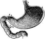
The inside of the stomach with the beginning of the intestines. At 3 is the left end and at 4 is the right end. At 1 is the opening of the gullet into the stomach. At 5 is a valve (the pylorus) which is sometimes shut, so as to prevent anything from passing from the stomach into the intestines. 8 indicates the inside lining of the stomach which produces gastric juices. 6 shows the duct from the liver.

The Stomach

The inside of the stomach with the beginning of the intestines. At 3 is the left end and at 4 is the…
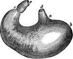
The stomach. Labels: d, lower end of the gullet; a, position of the cardiac aperture; b, the fundus; c, the pylorus; e, the commencement of the small intestine; along a, b, c, the great curvature; between the pylorus and d, the lesser curvature.

The Stomach

The stomach. Labels: d, lower end of the gullet; a, position of the cardiac aperture; b, the fundus;…
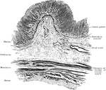
Transverse section of stomach (left end), showing general arrangement of coats.

Transverse Section of Stomach

Transverse section of stomach (left end), showing general arrangement of coats.
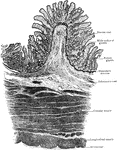
Transverse section of stomach, pyloric end; ruga is cut across, showing mucosa supported by core of submucous

Transverse Section of Stomach

Transverse section of stomach, pyloric end; ruga is cut across, showing mucosa supported by core of…
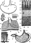
Views of the stomach. Labels: A. stomach (human). B. Same, anterior wall removed. C. Portion of stomach, pylorus and duodenum. D. Section through coats of stomach. E. Fundus of a cardiac gland. F. Epithelium from surface of stomach. G. Arterie sand veins of mucous membrane. w. Esophagus. 2. Duodenum. 3. Pylorus. $. Cardia. 5. Lesser curvature. 6. Greater curvature. 7. Anterior surface. 8. Pyloric valve. 9. Mucous membrane, with rugae. 10. Inner surface of gastric mucous membrane. 11. Mucous membrane of duodenum with villi. 12. Pyloric gastric glands. 13. Circular or deep muscular fibers. 17. Mucous membrane. 18. Muscularis mucosae. 19. Submucous coat. 20. Circular muscular layer. 21. Longitudinal muscular layer. 22. Serous coat. 23. Orifice of gland. 24. Fundus of gland.

Views of the Stomach

Views of the stomach. Labels: A. stomach (human). B. Same, anterior wall removed. C. Portion of stomach,…

Section through the liver and stomach, at the level of the xiphoid process.

Cross Section of the Trunk Through the Liver and Stomach

Section through the liver and stomach, at the level of the xiphoid process.
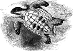
An imbricated turtle, flipped onto its back, revealing its underbelly.

Imbricated turtle, overturned

An imbricated turtle, flipped onto its back, revealing its underbelly.
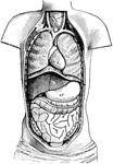
Diagram of the Body opened from the front to show the contents of the ventral cavity. Labels: d, diaphragm; h, heart; lu, lungs; st, stomach; li, liver; si, small intestines; c, large intestine

Ventral Cavity of the Body

Diagram of the Body opened from the front to show the contents of the ventral cavity. Labels: d, diaphragm;…