Earthworm AnatomyClipart ETC: An online service of Florida's Educational Technology Clearinghouse

Earthworm Anatomy

Earthworm AnatomyEarthworm AnatomyEarthworm Anatomy

To use any of the clipart images above (including the thumbnail image in the top left corner), just click and drag the picture to your desktop. You may also control-click (Mac) or right-click (Windows) and choose "Save file to disk" from the pop-up menu. Click here for help downloading and using clipart files.

Download TIFF* File

* A TIFF file is a high-resolution (240 dpi) format. TIFF files should be used only if you are printing the clipart. Do NOT use TIFF files for clipart that will displayed on a computer screen. Click here for help downloading and using TIFF files.

File Name: 69407_worms_earth
Description: 
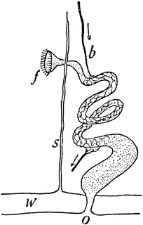
The earthworms are also known as megadriles, in the families Tubificidae, Lumbriculidae, and Enchytraeidae. Earthworms have a pair of kidneys to every segment, each consisting of a coiled tube wrapped in a mass of small blood vessels, and at its inner end communicating with the body cavity by means of a funnel-shaped opening. Here is a diagram of an earthworm kidney., showing (b) blood vessel, (f) funnel opening into body cavity, (o) outer opening, (s)septum, (w) body wall
Source: Winchell, Alexander Sketches of Creation (New York: Harper & Brothers, 1870) 74
Keywords: worm, earthworms, megadriles, Tubificidae, Lumbriculidae, Enchytraeidae

Copyright: 2009, Florida Center for Instructional Technology. See license.

Main MenuSite MapSearchLicenseClipart Help


Clipart ETC is a part of the Educational Technology Clearinghouse and is funded by various grants. Produced by the Florida Center for Instructional Technology, College of Education, University of South Florida. Email the project manager.


Clipart ETCEducational Technology Clearinghouse