Clipart tagged: ‘ankle’
Achillles tendon
"Tendons are white, glistening cords, or straps, which connect the muscles with the bones." —…
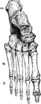
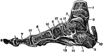
Bones of the Ankle and Foot
Bones of the Ankle and Foot. Labels: m, metatarsal bones; p, phalanges; ca, os calcis, or heel bone.
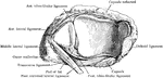
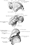
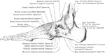
Ankle Joint and Foot
Vertical section of the ankle joint and foot. Labels: 1, tibia; 2, astragalus; 3, os calci; 4, scaphoides;…
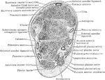
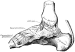
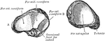
Section Through Ankle Joint
Coronal section through the left ankle joint, astragalus, and calcaneum.

Frontal Section of Foot and Ankle
Frontal section of the right ankle and foot. Viewed from in front.
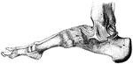
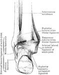
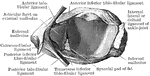
Ligaments of the Foot and Ankle
"The bones are fastened together, kept in place, and their movements limited, by tough and strong bands,…
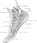
Sagittal Section of Foot and Ankle
Sagittal section of the foot and ankle passing through the great toe.
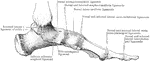
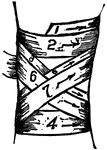
Outer Aspect of Foot Ligaments
Ligaments on outer aspect of ankle and on dorsum and outer aspects of foot.
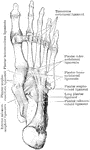
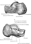
Longitudinal Section Through Foot
Longitudinal section through right foot in axis of first metatarsal bone.
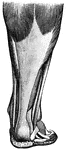
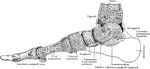
Muscles of the leg
"Muscles of the leg showing how they pass into tendons at the ankle." —Davison, 1910
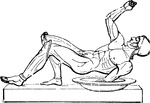
Fallen soldier
"Figure of a fallen warrior, represented among the sculptures now at Munich, belonging to the temple…
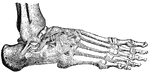
Tibia and Fibula
"The leg consists, like the forearm, of two bones. The larger, a strong, three-sided bone with…