Circulation in frog's foot under a microscope. Labels: A, walls of capillaries; B, tissue of web lying between the capillaries; C, cells of epidermis covering web (these are only shown in the right-hand and lower part of the field; in the other parts of the field the focus of the microscope lies below the epidermis); D, nuclei of these epidermic cells; E, pigment cells contracted, not partially expanded; F, red blood corpuscle (oval in the frog) passing along capillary -- nucleus not visible; G, another corpuscle squeezing its way through a capillary, the canal of which is smaller than its own transverse diameter; H, another bending as it slides round a corner; K, corpuscle in capillary seen through the epidermis; I, white blood corpuscle.

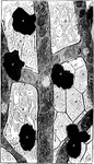
Circulation in a Frog's Foot

Circulation in frog's foot under a microscope. Labels: A, walls of capillaries; B, tissue of web lying…

The illustration exhibits the typical characters of the red blood cells in the main divisions of Vertbrata. The fractions are those of an inch, and represent the average diameter. In the case of the oval cells, only the long diameter is here given. It is remarkable, that although the size of the red blood cells varies so much in the different classes of the vertebrate kingdom, that of the white corpuscles remains comparatively uniform, and thus are, in some animals, much greater, in others much less than the red corpuscles existing side by side with them.

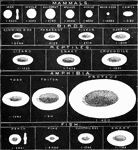
Red Blood Cells in Vertebrata

The illustration exhibits the typical characters of the red blood cells in the main divisions of Vertbrata.…