Clipart tagged: ‘"foot bone"’
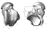
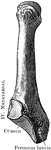
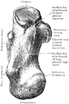
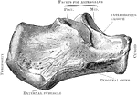
Astragalus
The right astragalus. A, Upper surface. B, Under surface. Labels: 1, groove for flex, long, hallucis;…
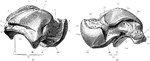
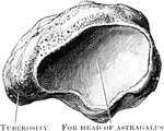
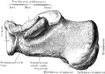
Astragalus
The right astragalus. A, As seen from the outer side. B, As seen from the inner side. Labels: 1, external…
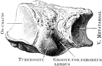
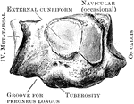
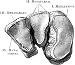
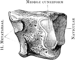
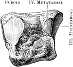
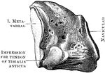
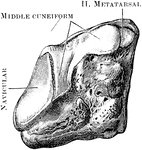
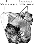
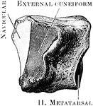
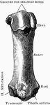
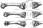
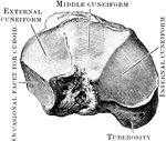
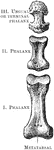
Metatarsal Bones
View of the bases and shafts of the second, third, and fourth metatarsal bones of the right foot.