"Head and Appendages of Honey-bee (Apis). a, Antenna or feeler. g, Epipharynx. mxp, Maxillary palp. pg, Opposite to galeae of 2nd maxillae (labium). mx, 1st maxilla. lp, Labial palp. l, Ligula or "tongue." b, Bouton or spoon of the ligula." — The Encyclopedia Britannica, 1910

Honey Bee

"Head and Appendages of Honey-bee (Apis). a, Antenna or feeler. g, Epipharynx. mxp, Maxillary palp.…
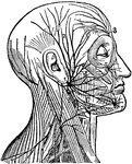
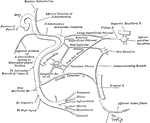
"(1) The facial nerve at its emergence from stylo-mastoid foramen; (2) temporal branches communicating with (3) the frontal branches of the fifth or trifacial nerve; (4) infraorbital branches communicating with (5) the infra-orbital branches of the fifth nerve; (6) maxillary branches communicating with (7) the mental branch of the fifth nerve; (8) cervico-facial branches; (9) the spinal accessory nerve giving off a branch to the trapezius muscles." — Winston's Encyclopedia, 1919

Facial Nerve

"(1) The facial nerve at its emergence from stylo-mastoid foramen; (2) temporal branches communicating…

Plan of the facial and intermediate nerves and their communication with other nerves.

Facial Nerves

Plan of the facial and intermediate nerves and their communication with other nerves.

"Fig. 62 Skull of common fowl, enlarged. from nature by Dr. R.W. Shufeldt, U.S.A. The names of bones and some other parts are printed, requiring no explanation; but observe the following points: The distinction of none of the bones composing the brain-case (the upper back expanded part) can be found in a mature skull. The brain is contained between the occipital, sphenoidals, squamosals, parietals and part of frontal; the ethmoidals belong to the same group of cranial bones proper. All other bones, excepting the three otic ear-bones, are bones of the face and jaws. The lower jaw, of five bones, is drawn detached; it articulates by the black surface marked articular with the prominence just above- the quadratic bone. Observe that from this quadrate a series of bones quadrato-jugal, jugal, maxillary-makes a slender rod running to the premaxillary; this is the zygoma, or jugal bar. Observe from the quadrate also another series, composed of pterygoid and palatine bones, to the premaxillary; this is the pterygo-palatine bar; it slides along a median fixed axis of the skull, the rostrum, which bears the loose vomer at its end. The under mandible, quadrate, pterygoid, and vomer are the only movable bones of this skull. But when the quadrate rocks back and forth, as it does by its upper joint, its lower end pulls and pushes upon the upper mandible, by means of the jugal and pterygo-palatine bars, setting the whole scaffolding of the upper jaw in motion. This motion hinges upon the elasticity of the bones of the forehead, at the thin place just where the reference-lines from the words "lacrymal" and "mesethmoid" cross each other. The dark oval space behind the quadrate is the external orifice of the ear; the parts in it to which the three reference-lines go are diagrammatic, not actual representations; thus, the quadrate articulates with a large pro-otic as well as with the squamosal. The great excavation at the middle of the figure, containing the cirlet of the unshaded bones, is the left orbital cavity, orbit, or socket of the eye. The mesethmoid includes most of the background of this cavity, shaded diagonally. The upper one of the two processes of bone extending into it from behind is post-frontal or sphenotic process; the under one (just over the quadrate) is the squamosal process. A bone not shown, the presphenoid, lies just in front of the oval black space over the end of basisphenoid. This black oval is the optic foramen, through which the nerve of sight passes from the brain-cavity to the eye. The black dot a little behind the optic foramen is the orifice of exit of a part of the trifacial nerve. The black mark under the letters "on" of the word "frontal" is the olfactory foramen, where the nerve of smell emerges from the brain-box to go to the nose. The nasal cavity is the black space behind nasal and covered by that bone, and in the oval blank before it. The parts of the beak covered by horn are only premaxillary, nasal, and dentary. The condyle articulates with the first cervical vertebra; just above it, not shown, is the foramen magnum, or great hole through which the spinal medulla, or main nervous cord, passes from the spinal column. The basioccipital is hidden, excepting its condyle; so is much of the basisphenoid. The prolongation forward of the basisphenoid, marked "rostrum," and bearing the vomer at its end, is the parasphenoid, as far as its thickened under border is concerned. Between the fore end of the pterygoid and the basisphenoidal rostrum, is the site of the basipterygoid process, by which the bones concerned articulate by smooth facets; further forward, the palatines ride freely upon the parasphenoidal rostrum. In any passerine bird , the vomer would be thick in front, and forked behind, riding like the palatine upon the rostrum. The palatine seems to run into the maxillary in this view; but it continues on to premaxillary. The maxillo-palatine is an important bone which cannot be seen in the figure because it extends horizontally into the paper from the maxillary about where the reference life "maxillary" goes to that bone. The general line from the condyle to the end of the vomer is the cranial axis, basis cranii, or base of the cranium. This skull is widest across the post-frontal; next most so across the bulge of the jugal bar." Elliot Coues, 1884

Skull of a Common Fowl

"Fig. 62 Skull of common fowl, enlarged. from nature by Dr. R.W. Shufeldt, U.S.A. The names of bones…

"Skull of Hystrix Eristata. t, temporal muscle; m, masseter. m', portion of masseter transmitted through the infra-orbital foramen, the superior maxillary nerve passing outwards between it and the maxillary bone." —The Encyclopedia Britannica, 1903

Hystrix Eristata

"Skull of Hystrix Eristata. t, temporal muscle; m, masseter. m', portion of masseter transmitted through…
Inferior surface of upper jaw at about birth.

Inferior Surface of Upper Jaw at Birth

Inferior surface of upper jaw at about birth.

Mesial surface of upper jaw at about birth.

Mesial Surface of Upper Jaw at Birth

Mesial surface of upper jaw at about birth.

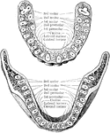
Horizontal section through both the upper and lower jaws to show the roots of the teeth. The sections were carried through the bones a short distance from the edge of their alveolar borders. The upper figure shows the upper teeth, the lower figure the lower teeth. Note the flattened roots of the lower incisors, the two root canals in the anterior root of each lower molar, and the confluence of the three roots of the upper wisdom teeth.

Jaw Showing Roots of Teeth

Horizontal section through both the upper and lower jaws to show the roots of the teeth. The sections…
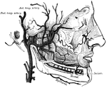
The internal maxillary artery and its branches.

Maxillary Artery

The internal maxillary artery and its branches.

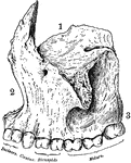
Superior maxillary bone. With it's fellow on the opposite side, it forms the whole of the upper jaw. Each bone assists in forming part of the floor of the orbit, the floor and outer wall of the nasal fossae, and the greater part of the roof of the mouth. Labels: 1, orbital surface; 2, facial surface; 3, alveolar process.

Human Maxillary (Upper Jaw) Bone

Superior maxillary bone. With it's fellow on the opposite side, it forms the whole of the upper jaw.…

Inferior Maxillary Bone (lower jaw). It is the largest and strongest bone in the face and serves for the reception of the lower teeth.

Human Maxillary (Upper Jaw) Bone

Inferior Maxillary Bone (lower jaw). It is the largest and strongest bone in the face and serves for…

Superior maxillary bone and the bony sinuses of the head.

Superior Maxillary Bone and Sinuses

Superior maxillary bone and the bony sinuses of the head.

Right superior maxillary bone, inner surface.

Superior Maxillary Bone

Right superior maxillary bone, inner surface.

Right infero-lateral view of the muscles of the maxillary space, the ramus and hyoid cornu are cut away. Labels: a, lingualis; b, hyo-glossus longus; c, ho-glossus brevis; d, genio-hyo-glossus; e, genio-hyoideus; f, stylo-hyoideus; g, hyoideus magnus; h, levator palati; i, tensor palati; k, pharyngostaphylinus; l, pterygo-pharyngeus; m, kerato-pharyngeus; n, hyo-pharyngeus; o, thyro-pharyngeus; p, crico-pharyngeus; q, esophagus.

Muscles of the Maxillary Space of a Horse

Right infero-lateral view of the muscles of the maxillary space, the ramus and hyoid cornu are cut away.…

"Skull of Polyodon. n, nasal cavity; sq, squamosal; mh, hyomandibular; sy, symplectic; pa, palate-pterygold; m, Meckelian cartiledge; mx, maxillary; d, dentary; h, hyoid; op, operculum; br, branchiostergal; s.cl, supraclavicular; p.cl, postclavicular; cl, clavicle; i.cl, infraclavicular." — Encyclopedia Britannica, 1893

Polyodon

"Skull of Polyodon. n, nasal cavity; sq, squamosal; mh, hyomandibular; sy, symplectic; pa, palate-pterygold;…

"Upper aspect of the primordial cranium, with the membrane-bones removed. An, angular; ao, anteorbital; Ar, articulary; B, basal; D, dentary; E, ethmoid; F, frontal; Ma, mastoid; Mp, metapterygold; Mx, maxillary; N, nasal; O, operculum; Oc, occipital; Pa, parietal; Pl, palatine; Pmx, premaxillary; po, postorbital; Prf, prefrontal; Pt, post-temporals; Ptf, postfrontal; Ptr, pterygold; Q, quadrate; S, suspensorium; So, suboperculum; Sp, sphenold; Spl, splenial; St, supratemporals; T, tympanic lamina; Tu, turbinal; v, vomer; x x, small ossicles; x' x', spiraculars." — Encyclopedia Britannica, 1893

Polypterus Skull

"Upper aspect of the primordial cranium, with the membrane-bones removed. An, angular; ao, anteorbital;…

"Lower aspect of the primordial cranium, with the membrane-bones removed. An, angular; ao, anteorbital; Ar, articulary; B, basal; D, dentary; E, ethmoid; F, frontal; Ma, mastoid; Mp, metapterygold; Mx, maxillary; N, nasal; O, operculum; Oc, occipital; Pa, parietal; Pl, palatine; Pmx, premaxillary; po, postorbital; Prf, prefrontal; Pt, post-temporals; Ptf, postfrontal; Ptr, pterygold; Q, quadrate; S, suspensorium; So, suboperculum; Sp, sphenold; Spl, splenial; St, supratemporals; T, tympanic lamina; Tu, turbinal; v, vomer; x x, small ossicles; x' x', spiraculars." — Encyclopedia Britannica, 1893

Polypterus Skull

"Lower aspect of the primordial cranium, with the membrane-bones removed. An, angular; ao, anteorbital;…

"Side view, with the membrane-bones. An, angular; ao, anteorbital; Ar, articulary; B, basal; D, dentary; E, ethmoid; F, frontal; Ma, mastoid; Mp, metapterygold; Mx, maxillary; N, nasal; O, operculum; Oc, occipital; Pa, parietal; Pl, palatine; Pmx, premaxillary; po, postorbital; Prf, prefrontal; Pt, post-temporals; Ptf, postfrontal; Ptr, pterygold; Q, quadrate; S, suspensorium; So, suboperculum; Sp, sphenold; Spl, splenial; St, supratemporals; T, tympanic lamina; Tu, turbinal; v, vomer; x x, small ossicles; x' x', spiraculars." — Encyclopedia Britannica, 1893

Polypterus Skull

"Side view, with the membrane-bones. An, angular; ao, anteorbital; Ar, articulary; B, basal; D, dentary;…

"Lower aspect of the skull, part of the bones being removed on the side. An, angular; ao, anteorbital; Ar, articulary; B, basal; D, dentary; E, ethmoid; F, frontal; Ma, mastoid; Mp, metapterygold; Mx, maxillary; N, nasal; O, operculum; Oc, occipital; Pa, parietal; Pl, palatine; Pmx, premaxillary; po, postorbital; Prf, prefrontal; Pt, post-temporals; Ptf, postfrontal; Ptr, pterygold; Q, quadrate; S, suspensorium; So, suboperculum; Sp, sphenold; Spl, splenial; St, supratemporals; T, tympanic lamina; Tu, turbinal; v, vomer; x x, small ossicles; x' x', spiraculars." — Encyclopedia Britannica, 1893

Polypterus Skull

"Lower aspect of the skull, part of the bones being removed on the side. An, angular; ao, anteorbital;…
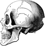
The skull. Labels: a, nasal bone; b, superior maxillary; c, inferior maxillary; d, occipital; e, temporal; f, parietal; g, frontal bone.

Human Skull

The skull. Labels: a, nasal bone; b, superior maxillary; c, inferior maxillary; d, occipital; e, temporal;…
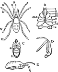
"Stylocellus sumatranus, one of the Opiliones; after Thorell. Enlarged. A, Dorsal view; I to VI, the six prosomatic appendages. B, Ventral view of the prosoma and of the first somite of the opisthosoma, with the appendages I to VI cut off at the base; a, tracheal stigma; mx, maxillary processes of the coxae of the 3rd pair of appendages; g, genital aperture. C, Ventral surface of the prosoma and opisthosoma; a, tracheal stigma; b, last somite. D, Lateral view of the 1st and 2nd pair of apendages. E, Lateral view of the whole body and two 1st appendages, showing the fusion of the dorsal elements of the prosoma into a single plate, and of those of the opisthosoma into an imperfectly segmented plate continuous with that of the prosoma." — The Encyclopedia Britannica, 1910

Stylocellus Sumatranus

"Stylocellus sumatranus, one of the Opiliones; after Thorell. Enlarged. A, Dorsal view; I to VI, the…
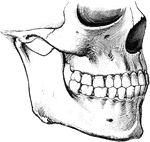
To show the relation of the upper to the lower teeth when the mouth is closed. The manner in which a tooth of one row usually strikes against two teeth of the opposite row, and the resulting interlocking of the teeth, is to be noted.

Teeth

To show the relation of the upper to the lower teeth when the mouth is closed. The manner in which a…
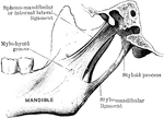
Internal lateral ligament of the temporo maxillary joint.

Ligament of the Temporo Maxillary Joint

Internal lateral ligament of the temporo maxillary joint.