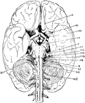
The base of the brain. The cerebral hemispheres are seen overlapping all the rest. Labels: I, olfactory lobes; II, optic tract passing to the optic chiasma from which the optic nerves proceed; III, the third nerve or motor oculi; IV, the fourth nerve or patheticus; V, the fifth nerve or trigminalis; VI, the sixth nerve or abducens; VII, the seventh or facial nerve; VIII, the auditory nerve; IX, the ninth or glossopharyngeal; X, the tenth or pneumogastric or vagus; XI, the spinal accessory; XII, the hypoglossal; ncI, the first cervical spinal nerve.

Base of the Brain

The base of the brain. The cerebral hemispheres are seen overlapping all the rest. Labels: I, olfactory…
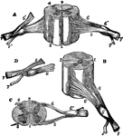
Views of section of cervical cord. Labels: A, anterior surface; B, right side; C, upper surface; D, nerve roots; 1, anterior median fissure; 2, posterior median fissure; 3, anterior lateral depression; 4, posterior lateral groove; 5, anterior roots; 6, posterior roots; 6', ganglion; 7, spinal nerve; 7', posterior branch.

Sections of Cervical Spinal Cord

Views of section of cervical cord. Labels: A, anterior surface; B, right side; C, upper surface; D,…

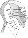
Cutaneous nerve areas of the head and neck.

Surface Areas of Nerves of the Head and Neck

Cutaneous nerve areas of the head and neck.

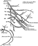
Diagrammatic view from before of the spinal cord and medulla oblongata, including the roots of the spinal and some of the cranial nerves, and on one side of the gangliated chain of the sympathetic. The spinal nerves are enumerated in order on the right side of the figure. Labels: Br, brachial plexus; Cr, anterior crural, O, obturator, and Ss, great sciatic nerves, coming off from lumbo-sacral plexus; x, x, filum terminale; a,b,c, superior, middle, and inferior cervical ganglia of the sympathetic, the last united with the first thoracic, d; d', the eleventh thoracic ganglion; l, the twelfth thoracic (or first lumbar); below ss, the chain of sacral ganglia.

Spinal Cord

Diagrammatic view from before of the spinal cord and medulla oblongata, including the roots of the spinal…
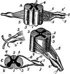
Diagrams of spinal cord and nerve roots. Labels: A, a small portion of the cord seen from the ventral side; B, the same seen laterally; C, a cross-section of the cord; D, the two roots of a spinal nerve; 1, anterior (ventral) fissure; 2, posterior (dorsal) fissure; 3, surface groove along the line of attachment of the anterior nerve roots; 4, line of origin of the posterior roots; 5, anterior root filaments of a spinal nerve; 6, posterior root filaments; 6', ganglion of the posterior root; 7, 7', the first two divisions of the nerve trunk after its formation by the union of the two roots.

Spinal Cord and Nerve Roots

Diagrams of spinal cord and nerve roots. Labels: A, a small portion of the cord seen from the ventral…
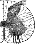
A thin transverse section of half the spinal cord magnified about 10 diameters. Labels: 1, anterior fissure; 2, posterior fissure; 3, central canal ;8, pia mater enveloping the cord; 6,7, bands of pia mater penetrating the cord and supporting its nerve elements; 9, a posterior root; 10, bundles of an anterior root; a,b,c,d,e, groups of nerve cells in the gray matter.

Spinal Cord Section

A thin transverse section of half the spinal cord magnified about 10 diameters. Labels: 1, anterior…

Illustrating the functions of the spinal nerves. Divided at a. -- Irritated at 1: pain. Irritated at 2: muscular contraction. Nerve trunks transmit stimuli which give rise to sensation, as, for example, sensations of pain (sensory stimuli).

Spinal Nerves

Illustrating the functions of the spinal nerves. Divided at a. -- Irritated at 1: pain. Irritated at…

The spinal nerves.

The Spinal Nerves

The spinal nerves.

The brachial plexus of the spinal nerves.

The Brachial Plexus of the Spinal Nerves

The brachial plexus of the spinal nerves.

The brachial plexus of the spinal nerves, and nerves of the upper extremity.

The Brachial Plexus of the Spinal Nerves

The brachial plexus of the spinal nerves, and nerves of the upper extremity.

The cervical plexus of the spinal nerves.

The Cervical Plexus of the Spinal Nerves

The cervical plexus of the spinal nerves.

The lumbar and sacral plexuses of the spinal nerves.

The Lumbar and Sacral Plexuses of the Spinal Nerves

The lumbar and sacral plexuses of the spinal nerves.

The lumbar and sacral plexuses of the spinal nerves, showing the distribution of nerve branches.

The Lumbar and Sacral Plexuses of the Spinal Nerves

The lumbar and sacral plexuses of the spinal nerves, showing the distribution of nerve branches.

Illustrating the functions of the roots of the spinal nerves. Labels: a, ventral root; p, dorsal root. Divided at a. -- Irritated at 1: no result. Irritated at 2: contraction of muscles supplied with fibers from the root. Divided at p. -- Irritated at 3: no result. Irritated at 4: pain produced.

Roots of Spinal Nerves

Illustrating the functions of the roots of the spinal nerves. Labels: a, ventral root; p, dorsal root.…