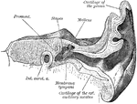
Transverse section of external auditory canal and tympanum. Left side.

Auditory Canal and Tympanum

Transverse section of external auditory canal and tympanum. Left side.

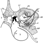
"Diagram showing the ear and related parts in a young cat. P., Pinna; Sq., squamosal: E.A.M., external auditory meatus; T., tympanum; M., malleus; I., incus; St., stapes abutting onforamen ovale; B., bulla of tympanic bone; Se., a septum in the bulla; E.T., eustachian tube leading from the tymmpanic cavity to the back of the mouth; B.O., basi-occipital; C., cochlea; S., sacculus; U., utriculus; D.E., ductus endolymphaticus; N., auditory nerve; S.C., semi-circular canal; PE., periotic bone." -Thomson, 1916

Cat Ear

"Diagram showing the ear and related parts in a young cat. P., Pinna; Sq., squamosal: E.A.M., external…
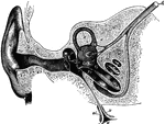
Semi-diagrammatic section through the right ear. Labels: M, concha; G, the external auditory canal; T, tympanic, or drum-membrane (ear drum); P, tympanum, or middle ear; o, oval window; r, round window. Extending from T to o is seen the chain of the tympanic bones; R, Eustachian tube; V, B, S, bony labyrinth; V, vestibule; B, semicircular canal; S, cochlea; b, l, v, membranous labyrinth in semi-circular canal and in vestibule. A, auditory nerve dividing into branches for vestibule, semicircular canal, cochlea.

Ear and Auditory Canal

Semi-diagrammatic section through the right ear. Labels: M, concha; G, the external auditory canal;…
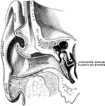
Vertical section through the external auditory canal and tympanum, passing in front of the fenestra ovalis.

Ear Showing Auditory Canal and Tympanum

Vertical section through the external auditory canal and tympanum, passing in front of the fenestra…
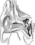
Vertical section through the external auditory meatus and tympanum, passing in front of the fenestra ovalis.

Ear Showing External Auditory Meatus

Vertical section through the external auditory meatus and tympanum, passing in front of the fenestra…

Bones of the ear: Malleus, Incus and Orbiculare, Stapes. The bones of the area are connected with each other in the drum and serve to transmit vibrations of the membrane of the tympanum to the internal ear. They are very hard and brittle, with no cartilage on their articular surfaces like other bones.

Bones of the Ear

Bones of the ear: Malleus, Incus and Orbiculare, Stapes. The bones of the area are connected with each…
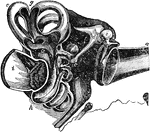
General sectional view of the structure of the ear. Labels: a, the meatus auditorius externus; b, the tympanum; c, the malleus; d, the incus, e, the os orbiculare; f, the stapes; g, the semicircular canals; h, the cochlea; i, the meatus auditorius internus; k, the eustachian tube.

Sectional View of the Ear

General sectional view of the structure of the ear. Labels: a, the meatus auditorius externus; b, the…
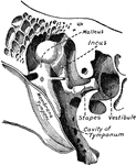
Chains of small bones and their ligaments, seen from the front in a vertical, transverse section of the tympanum.

Small Bones and Ligaments of the Ear

Chains of small bones and their ligaments, seen from the front in a vertical, transverse section of…
The Tympanum Medusa Head is found in the arch of the entrance of the Royal Palace of Tuileries in Paris, France.

Tympanum Medusa Head

The Tympanum Medusa Head is found in the arch of the entrance of the Royal Palace of Tuileries in Paris,…
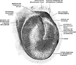
The membrana tympani separates the cavity of the tympanum from the bottom on the external canal. Shown is the right membrana tympani, viewed from the outside, from in front, and from below.

Membrana Tympani

The membrana tympani separates the cavity of the tympanum from the bottom on the external canal. Shown…
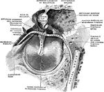
The membrana tympani separates the cavity of the tympanum from the bottom on the external canal. Shown is the right membrana tympani with the hammer and the chorda tympani, viewed from within, from behind, and from above.

Membrana Tympani

The membrana tympani separates the cavity of the tympanum from the bottom on the external canal. Shown…

Diagram to illustrate the action of the ossicles of the middle ear in the conduction of sound to the internal ear.

Ossicles of the Middle Ear

Diagram to illustrate the action of the ossicles of the middle ear in the conduction of sound to the…
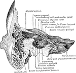
Section through the petrous and mastoid portions of the temporal bone, showing the communication of the cavity of the tympanum with the mastoid antrum.

Section Through Temporal Bone

Section through the petrous and mastoid portions of the temporal bone, showing the communication of…
Section through left temporal bone, showing inner wall of tympanic cavity.

Tympanic Cavity

Section through left temporal bone, showing inner wall of tympanic cavity.

Left tympanic membrane (as viewed from the external auditory meatus).

Tympanic Membrane

Left tympanic membrane (as viewed from the external auditory meatus).

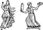
"Tympanum, a small drum carried in the hand. Of these, some resembled in all respects a modern tambourine with bells. Others presented a flat circular disk on the upper surface and swelled out beneath like a kettle-drum. Both forms are represented in the cuts below. Tympana were covered with the hides of oxen, or of asses; were beaten with a stick, or with the hand, and were much employed in all wild enthusiastic religious rites, especially the orgies of Bacchus and Cybele." &mdash Smith; 1873

Tympanum

"Tympanum, a small drum carried in the hand. Of these, some resembled in all respects a modern tambourine…
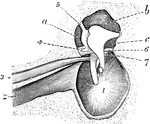
Interior view of the tympanum, with membrana tympani and bones in natural position. 1, Membrana tympani; 2, Eustachian tube; 3, tensor tympani muscle; 4, lig. mallei super; 6, corda-tympani nerve; a, b, and c, sinuses about ossicula.

Tympanum

Interior view of the tympanum, with membrana tympani and bones in natural position. 1, Membrana tympani;…