Clipart tagged: ‘vertebrae’
A Cervical Vertebra
A cervical vertebra. Labels: Frt, vertebral foramen; Pai, anterior articular process; R, rudimentary…
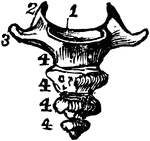
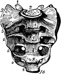
Human Cervical Vertebra Bone
A cervical vertebra of the spine, inferior surface. Labels: 1, spinous process, slightly bifid; 4, transverse…
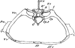
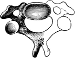
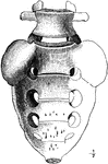
Crocodile Thoracic Region
"Segment of Endoskeleton from Thoracic Region of Crocodile. C, centrum of a vertebra, over which rises…
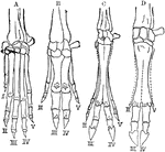
Hands of Vertebrates
A comparison of vertebrate hands. A, hand or anterior foot of the dog; B, that of the hog; C, that of…
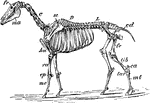
Horse Skeleton
"Skeleton of Horse (Equus caballus). fr, frontal bone; C, cervical vertebrae; D, dorsal vertebrae; L,…
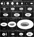
Red Blood Cells in Vertebrata
The illustration exhibits the typical characters of the red blood cells in the main divisions of Vertbrata.…
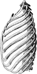
The Ribs
The ribs of the left side, with the dorsal and two lumbar vertebrae, the rib cartilages, and the sternum.
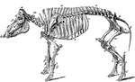
Skeleton of a Dog
The skeleton of the dog. Axial skeleton. The skull. Cranial bones- a, occipital, 1; b, parietal, 2;…
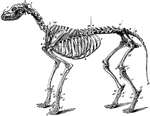
Skeleton of a Hog
Skeleton of the hog. Axial skeleton. The skull. Cranial bones- a, occipital, 1; b, parietal, 2; d, frontal,…
Spinal Accessory Nerve
Scheme of the origin, connection, and distribution of the spinal accessory nerve. Labels: Sp.Acc, spinal…
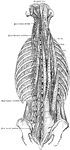
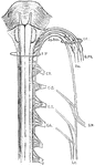
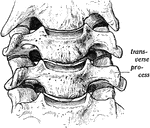
Sacral Region of Spinal Canal
The conus and medullaris and the filum terminale exposed within the spinal canal.
Spinal Column
Side view of the spinal column, with the vertebrae numbered: C1-7, cervical vertebrae; D1-12, dorsal…
Spinal Column
Lateral view of the spinal column. Labels: 1, atlas; 2, dentata 3, seventh cervical vertebra; 4, twelfth…
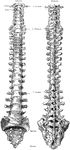
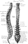
The Spinal Column
View of the entire spinal column. The bodies of the vertebrae are in the front with the spinous processes…
Human Spinal Column
Side view of spinal column, without sacrum and coccyx. Labels: 1 to 7, cervical vertebrae; 8 to 19,…
Side View of the Spinal Column
Side view of the spinal column. Labels: C 1-7, cervical; D 1-12, dorsal; L 1-15 lumbar; S 1, sacrum;…
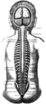
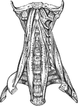
The Position of the Spinal Cord and Spinal Nerves in the Spinal Canal
The skull and spinal canal of a child from behind with the Dura Mater slit open and ribs with the transverse…
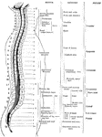
Distribution of the Spinal Nerves
Table giving approximate areas of distribution of the different spinal nerves with a diagram showing…
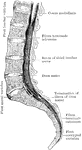
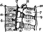
Spine
"The spine, sawn in two lengthwise, showing the spinal canal and the holes between the vertebrae, where…
The Spine
The spine showing the seven vertebrae of the neck, cervical; the twelve of the back, dorsal; the five…
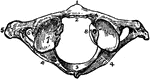
The Atlas Vertebra
The atlas, the uppermost vertebra of the spinal column. Labels: 1, anterior tubercle; 2, articular face…
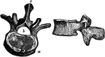
Two Views of a Vertebra
Two views of vertebra. 24 vertebrae make up the spinal column. On the left figure, a is the…
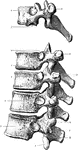
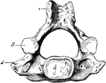
Thoracic Vertebrae
First, ninth, tenth, eleventh, and twelfth thoracic vertebrae from the left side. 1, Inferior articular…
Lateral and Posterior View of the Vertebral Column
Lateral and posterior views of the vertebral column.