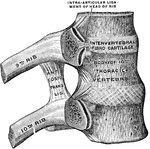
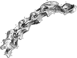
Ribs and corresponding vertebral bodies in their ligaments, viewed from the right.

Ribs and Corresponding Vertebral Bodies

Ribs and corresponding vertebral bodies in their ligaments, viewed from the right.

The conus and medullaris and the filum terminale exposed within the spinal canal.

Sacral Region of Spinal Canal

The conus and medullaris and the filum terminale exposed within the spinal canal.

Side view of the spinal column, with the vertebrae numbered: C1-7, cervical vertebrae; D1-12, dorsal vertebrae; L1-4, lumbar vertebrae; S1, sacrum; Co1-4, coccyx.

Spinal Column

Side view of the spinal column, with the vertebrae numbered: C1-7, cervical vertebrae; D1-12, dorsal…

Lateral view of the vertebral column.

Spinal Column

Lateral view of the vertebral column.

Diagrammatic representation of the roots and ganglia of the spinal nerves, showing their position in relation to the spinal column. The nerves are shown as thick black lines on the left side.

Spinal Nerves

Diagrammatic representation of the roots and ganglia of the spinal nerves, showing their position in…
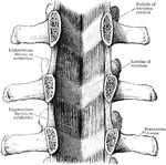
Ligamenta subflava as seen from the front after removal of he bodies as the vertebrae by sawing through the pedicles.

Ligamenta Subflava of the Spine

Ligamenta subflava as seen from the front after removal of he bodies as the vertebrae by sawing through…

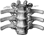
Anterior common ligament of the vertebral column, and the costo vertebral joints as seen from in front.
Ligaments of the Spine

Anterior common ligament of the vertebral column, and the costo vertebral joints as seen from in front.

Posterior common ligament of the vertebral column.

Ligaments of the Spine

Posterior common ligament of the vertebral column.

Mesial section through a portion of the lumbar part of the spine.

Mesial Section of the Spine

Mesial section through a portion of the lumbar part of the spine.

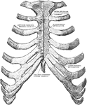
Sternum and ribs with ligaments, from in front. In the right half of the figure the most anterior layer has been removed and the joint cavities have been opened; the parts are separated somewhat from one another on the left side.

Sternum and Ribs

Sternum and ribs with ligaments, from in front. In the right half of the figure the most anterior layer…
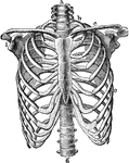
The skeleton of the thorax. Labels: a, g, vertebral column; b, first rib; c, clavicle; e, seventh rib; i, glenoid fossa.

Thorax Skeleton

The skeleton of the thorax. Labels: a, g, vertebral column; b, first rib; c, clavicle; e, seventh rib;…
Vertebral column with ligaments, from in front.

Vertebral Column with Ligaments

Vertebral column with ligaments, from in front.

The vertebral column from behind.

Back View of Vertebral Column

The vertebral column from behind.

Right lateral view of the cervical portion of the vertebral column of a horse. 1 to 7, the seven segments; 1, the atlas-a, its wing-S, internal foramen of alar gutter-9, posterior foramen of wing; 2, the dentata-11, neural spine; 12, transverse process; 13, centrum; 14, foramen corresponding to intervertebral foramen; 10, atlo-axoid space; the odontoid process is seen below the figures; 15, zygapophyses, or olbique processes; 16, centra, or bodies, in situ- 17, hypapophyses, or inferior processes-b, transverse processes-c, trifid transverse process of sixth vertebra-d, transverse process of seventh.

Horse Vertebral Column

Right lateral view of the cervical portion of the vertebral column of a horse. 1 to 7, the seven segments;…

The spinal column, right lateral view and dorsal view.

Lateral and Dorsal View of the Vertebral Column

The spinal column, right lateral view and dorsal view.

Lateral and posterior views of the vertebral column.

Lateral and Posterior View of the Vertebral Column

Lateral and posterior views of the vertebral column.

The vertebral column from the left side.

Side View of Vertebral Column

The vertebral column from the left side.