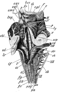
Medulla

Dorsal or posterior view of the medulla, fourth ventricle, and mesencephalon. Labels: p.n., line of the posterior roots of the spinal nerves; p.m.f., posterior median fissure; f.g., funiculus gracilis; cl., its clava; f.c., funiculus cuneatus; f.R., funiculus of Rolando; r.b., restiform body; c.s., calamus scriptorius; l, section of ligula or taenia; part of choroid plexus is seen beneath t; l.r., lateral recess of the ventricle; str., striae acusticae; i.f., inferior fossa; s.f., posterior fossa; between it and the median sulcus is the fasciculus teres; cbl., cut surface of the cerebellar hemisphere; nd., central or gray matter; s.m.v., superior medullary velum; lng., ligula; s.c.p., superior cerebellar peduncle cut longitudinally; cr., combined section of the three cerebellar peduncles; c.q.s., c.q.i., corpora quadrigemina (superior and inferior); fr., fraenulum; f; f., fibers of the fillet seen on the surface of the tegmentum; c, crusti; l.g., lateral groove; c.g.i, corpus geniculum internus; th., posterior part of the thalamus; p., pineal body. The Roman numbers indicate the corresponding cranial nerves.
Galleries
Human Central Nervous SystemSource
Baker, W. Morrant & Harris, Vincent Dormer Kirkes' Hand-book of Physiology, 13th ed. (Philadelphia: P. Blakiston's Son & Co., 1892) 607
Downloads
1523×2400, 1.2 MiB
649×1024, 184.0 KiB
406×640, 83.1 KiB
203×320, 24.4 KiB