Eye of Fetus of Four Weeks
| View Cart ⇗ | Info
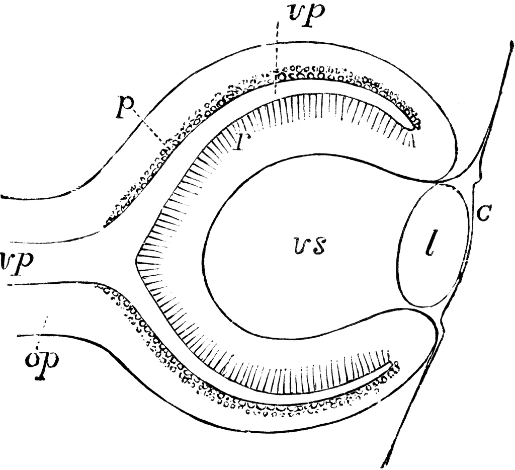
Diagrammatic sketch of a vertical longitudinal section through the eyeball of a human fetus of four weeks. The section is a little to the side, so as to avoid passing through the ocular cleft; c, the cuticle where it becomes later the corneal epithelium; l, the lens; op, optic nerve formed by the pedicle of the primary optic vesicle; vp, primary medullary cavity or optic vesicle; p, the pigment layer of the retina; r, the inner wall forming the retina proper; vs, secondary optic vesicle containing the rudiment of the vitreous humour.
Keywords
eyeSource
Baker, W. Morrant & Harris, Vincent Dormer Kirkes' Hand-book of Physiology, 13th ed. (Philadelphia: P. Blakiston's Son & Co., 1892) 837
Downloads
2400×2191, 483.1 KiB
1024×934, 71.2 KiB
640×584, 37.4 KiB
320×292, 14.8 KiB