This human anatomy ClipArt gallery offers 69 illustrations of human embryonic and fetal development, including external and dissected views of general development and development of specific systems progressing from fertilization to birth. Also includes views of the uterus and placenta as the embryo or fetus develops.
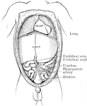
The abdominal and thoracic viscera of a five months fetus. The large liver and large size if its left lobe at this age should be noted.

Abdomen of Fetus

The abdominal and thoracic viscera of a five months fetus. The large liver and large size if its left…
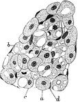
A lobule of developing adipose tissue from an eight month fetus. Labels: a, Spherical or, from pressure, polyhedral cells with large central nucleus surrounded by a finely reticulated substance staining uniformly with haematoxylin. b, Similar cells with spaces from which the fat has been removed by oil and cloves. c, Similar cells showing how the nucleus with enclosing protoplasm is being pressed towards periphery. d, Nucleus of endothelium of investing capillaries.

Developing Adipose Tissue in Fetus

A lobule of developing adipose tissue from an eight month fetus. Labels: a, Spherical or, from pressure,…
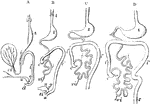
Outlines of the form and position of the alimentary canal in successive stages of its development. A, alimentary canal, in an embryo of four weeks; B, at six weeks; C, at eight weeks; D, at ten weeks; l, the primitive lungs connected with the pharynx; s, the stomach; d, duodenum; i, the small intestine; i', the large intestine; c, the caecum and vermiform appendage; r, the rectum; cl, in A, the cloaca; a, in B, the anus distinct from s i, the sinus uro-genitalis; v, the yolk sac; vi, the vitellointestinal duct; u, the urinary bladder and urachus leading to the allantois; g, genital ducts.

Development of the Alimentary Canal

Outlines of the form and position of the alimentary canal in successive stages of its development. A,…

The anal cavity and lower part of the rectum in the fetus. Left figure is four month. Middle figure is six months. Right figure is 9 months. In each the anal canal is distinctly marked off from the rectum proper; the columns of Morgagni and the rectal valves are distinct.

Development of the Anal Cavity

The anal cavity and lower part of the rectum in the fetus. Left figure is four month. Middle figure…
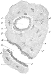
Transverse section of femur of a human embryo about eleven weeks old. Labels: a, rudimentary Haversian canal in cross sections; b, in longitudinal section; c, osteoblast; d, newly formed osseous substance of a lighter color; e, that of greater age; f, lacunae with their cells; g, a cell still united to an osteoblast.

Formation of Compact Bone in a Human

Transverse section of femur of a human embryo about eleven weeks old. Labels: a, rudimentary Haversian…
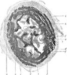
Cross section of developing bone of human fetus of four months. Labels: a, periostem; b, boundary between endochondral and periosteal bone; c, perichondral bone; d, remains of area of calcification; e, endochondral bone; f, f', blood vessels; g, g', developing Haversian spaces; h, marrow; i, blood vessels.

Developing Bone

Cross section of developing bone of human fetus of four months. Labels: a, periostem; b, boundary between…

Human fetus in the third month of development, with the brain and spinal cord exposed from behind.

Brain and Spinal Cord of Fetus

Human fetus in the third month of development, with the brain and spinal cord exposed from behind.

Side view of fetal brain at six months, showing commencement of formation of the principal fissures and convolutions. F, frontal lobe; P, parietal; O, occipital; T, temporal; a, commencing frontal convolutions; s, Sylvian fissure; s', its anterior division; c, within it the central lobe or island of Reil; r, fissure of Rolando; p, perpendicular fissure.

Fetal Brain at Six Months

Side view of fetal brain at six months, showing commencement of formation of the principal fissures…
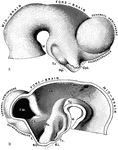
The brain of a human embryo in the fifth week. A, Brain as seen in profile. B, Mesial section through the same brain. Labels: M, mammillary eminence, Tc, tuber cinereum; Hp, Hypophysis (pituitary diverticulum from buccal cavity); Opt, optic stalk; TH, optic thalamus; Tg, tegmental part of mesencephalon; Ps, pars subtalamica; Cs, corpus striatum; FM, foramen of Monro; L, lamina terminalis; RO, recessus opticus; RI, recessus infundibuli.

Brain of Embryo

The brain of a human embryo in the fifth week. A, Brain as seen in profile. B, Mesial section through…
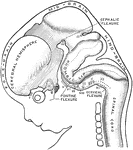
Profile view of brain of a human embryo of ten weeks. The various cranial nerves are indicated by numerals. A, Cerebral diverticulum of pituitary body. B, Buccal diverticulum of pituitary body.

Brain of Embryo

Profile view of brain of a human embryo of ten weeks. The various cranial nerves are indicated by numerals.…

The left cerebral hemisphere, from a fetus in the early part of the seventh month of development. Labels: p.c.s., sulcus praecentralis superior; p.c.i., sulcus praecentralis inferior; r1, lower part of Rolandic fissure; r2, upper part of Rolandic fissure; p1, inferior postcentral sulcus; p3, ramus horizontalis; p4, ramus occipitalis; e.p., external perpendicular fissure; t1, parallel sulcus; S, Sylian fossa; F.P., fronto-parietal wall; F.,frontal wall, O., orbital wall.

Brain of Fetus

The left cerebral hemisphere, from a fetus in the early part of the seventh month of development. Labels:…

Two stages in the development of the human brain. A. Brain of an human embryo of the third week. B. Brain of an embryo of five weeks.

Development of Human Brain

Two stages in the development of the human brain. A. Brain of an human embryo of the third week. B.…
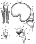
Early stages in development of human brain. 1, 2, 3, are from an embryo about seven weeks old; 4, about three months old. m, middle cerebral vesicle (mesencephalon); c, cerebellum; m o, medulla oblongata; i, thalamencephalon; h, hemispheres; i', infundibulum; Fig. 3 shows the several curves which occur in the course of development; Fig. 4 is a lateral view, showing the great enlargement of the cerebral hemispheres which have covered in the thalami, leaving the optic lobes, m, uncovered.

Development of the Brain

Early stages in development of human brain. 1, 2, 3, are from an embryo about seven weeks old; 4, about…
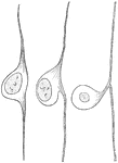
Three stages in the development of a cell from a spinal ganglion.

Development of Cell from Spinal Ganglion

Three stages in the development of a cell from a spinal ganglion.
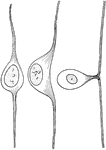
Three stages in the development of a cell from a spinal ganglion.

Development of a Cell from the Spinal Ganglion

Three stages in the development of a cell from a spinal ganglion.

Very soon after the entrance of the ovum into the uterus, in the human subject, the outer surface of the chorion is found beset with fine processes, the so-called chorion villi, which give it a rough and shaggy appearance. a, chorion with villi. The villi are shown to be best developed in the part of the chorion to which the allantois is extending, this portion ultimately becomes the placenta. ; b, space between the two layers of the amnion; c, amniotic cavity; d, situation of the intestine, showing its connection with the umbilical vesicle; e, umbilical vesicle; f, situation of heart and vessels; g, allantois.

Chorion Villi

Very soon after the entrance of the ovum into the uterus, in the human subject, the outer surface of…
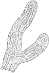
Very soon after the entrance of the ovum into the uterus, in the human subject, the outer surface of the chorion is found beset with fine processes, the so-called chorion villi, which give it a rough and shaggy appearance. Shown is a magnified view of chorion villi.

Magnified View of Chorion Villi

Very soon after the entrance of the ovum into the uterus, in the human subject, the outer surface of…

Diagram of fetal circulation.

Diagram of Fetal Circulation

Diagram of fetal circulation.

Plan of fetal circulation. In this plan, the figured arrows represent the kind of blood, as well as the direction which it takes in the vessels. Thus, arterial blood is figured ..... ; venous blood ----; mixed (arterial and venous) -...- .

Diagram of Fetal Circulation

Plan of fetal circulation. In this plan, the figured arrows represent the kind of blood, as well as…

Corpus lutea of different periods. B, corpus luteum of about the sixth week after impregnation, showing its plicated form at that period. 1, substance of the ovary; 2, substance of the corpus luteum; 3, a grayish coagulum in its cavity. A, corpus luteum two days after delivery; D, in the twelfth week after delivery.

Corpus Lutea of Different Periods

Corpus lutea of different periods. B, corpus luteum of about the sixth week after impregnation, showing…
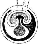
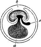
Diagram of fecundated egg. a, umbilical vesicle; b, amniotic cavity; c, allantois.

Fecundated Egg

Diagram of fecundated egg. a, umbilical vesicle; b, amniotic cavity; c, allantois.

Fecundated egg with allantois nearly complete. Labels: a, inner layer of amniotic fold; b, outer layer; c, point where the amniotic folds come in contact. The allantois is seen penetrating between the outer and inner layers of the amniotic folds.

Fecundated Egg

Fecundated egg with allantois nearly complete. Labels: a, inner layer of amniotic fold; b, outer layer;…
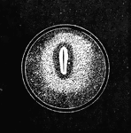
Impregnated egg, with commencement of formation of embryo; showing the area germinativa or embryonic spot, the area pellucida, and the primitive groove or trace.

Impregnated Egg

Impregnated egg, with commencement of formation of embryo; showing the area germinativa or embryonic…

A human embryo of the fourth week. I, the chorion; 3, part of the amnion; 4, umbilical vesicle with its long pedicle passing into the abdomen; 7, the heart; 8, the liver; 9, the visceral arch destined to form the lower jaw, beneath which are two other visceral arches separated by the branchial clefts; 10, rudiment of the upper extremity, 11, that of the lower extremity; 12, the umbilical cord; 15, the eye; 16, the ear; 17, cerebral hemispheres; 18, optic lobes, corpora quadrigemina.

Embryo at Fourth Week

A human embryo of the fourth week. I, the chorion; 3, part of the amnion; 4, umbilical vesicle with…

Human embryo of fifth week with umbilical vesicle. The human umbilical vesicle never exceeds the size of a small pea.

Embryo of Fifth Week

Human embryo of fifth week with umbilical vesicle. The human umbilical vesicle never exceeds the size…
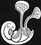
Diagram of young embryo and its vessels, showing course of circulation in the umbilical vesicle; and also that of the allantois (near the caudal extremity), which is just commencing.

Embryo Showing Course of Circulation

Diagram of young embryo and its vessels, showing course of circulation in the umbilical vesicle; and…
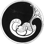
Diagram of embryo and its vessels, showing course of circulation. The large umbilical arteries are seen passing out in the placenta.

Embryo Showing Course of Circulation

Diagram of embryo and its vessels, showing course of circulation. The large umbilical arteries are seen…
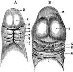
A, Magnified view of the head and neck of a human embryo of three weeks. Labels: 1, anterior cerebral vesicle or cerebrum; 2, middle cerebral vesicle; 3, middle or frontonasal process; 4, superior maxillary process, or first visceral arch, and below it the first cleft; 7, 8, 9, second, third, and fourth arches and clefts. B, Anterior view of the head of a human fetus of about the fifth week. 1, 2, 3, 5, the same parts as in A; 4, the external nasal or lateral frontal process: 6, the superior maxillary process; 7, the lower jaw; X, the tongue; 8, first branchial cleft becoming the meatus auditorius externus.

Head of an Embryo

A, Magnified view of the head and neck of a human embryo of three weeks. Labels: 1, anterior cerebral…
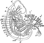
"Early Human Embryo, giving diagrammatically the principal vessels antecedent to the establishment of the regular fetal circulation. H, heart; P, lungs; L, liver; T A, the aortic trunk or cardiac aorta; c, c', c", common, external, and internal carotids; s, subclavian artery; v, vertebral artery; 1, 2, 3, 4, 5, the aortic arches (the persistent left aortic arch hidden); A, subvertebral aorta; o, o', omphalomeseraic artery and vein, to and from U, the umbilical vesicle with its vitelline duct, dv; u, u, the two hypogastric or umbilical arteries, with the ramifications, u", u", in the placenta; u', umbilical vein; vh, hepatic vein; cv, inferior vena cava; vil, iliac veins; az, an azygous vein; vc, a posterior cardinal vein; vi, innominate vein; vp, portal vein; Dv, the ductus venosus; DC, a ductus Cuvieri. The anterior cardinal vein is seen beginning in the head and running down to the ductus Cuvieri, on the under side of the numbers 1, 2, 3, 4, 5." -Whitney, 1911

Human Embryo

"Early Human Embryo, giving diagrammatically the principal vessels antecedent to the establishment of…

Diagram of a longitudinal section of an embryo, showing the different areas of the blastodermic vesicle. Labels: a, pericardium; b, bucco-pharyngeal; c, ectoderm; d, entoderm; e,placental area.

Longitudinal Section of an Embryo

Diagram of a longitudinal section of an embryo, showing the different areas of the blastodermic vesicle.…
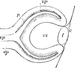
Diagrammatic sketch of a vertical longitudinal section through the eyeball of a human fetus of four weeks. The section is a little to the side, so as to avoid passing through the ocular cleft; c, the cuticle where it becomes later the corneal epithelium; l, the lens; op, optic nerve formed by the pedicle of the primary optic vesicle; vp, primary medullary cavity or optic vesicle; p, the pigment layer of the retina; r, the inner wall forming the retina proper; vs, secondary optic vesicle containing the rudiment of the vitreous humour.

Eye of Fetus of Four Weeks

Diagrammatic sketch of a vertical longitudinal section through the eyeball of a human fetus of four…
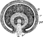
Transverse vertical section of the eyeball of a human embryo of four weeks. The anterior half of the section is represented: pr, the remains of the cavity of the primary optic vesicle; p, the inner part of the outer layer forming the retinal pigment; r, the thickened inner part giving rise to the columnar and other structures of the retina; v, the commencing vitreous humour within the secondary optic vesicle; v', the ocular cleft through which the loop of the central blood vessel, a, projects from below; l, the lends with a central cavity.

Eye of Fetus of Four Weeks

Transverse vertical section of the eyeball of a human embryo of four weeks. The anterior half of the…
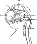
"Diagram of head and brain of human foetus six weeks old (heavy boundaries). The dotted line indicates the outline of the brain of a foetus three months old. Note thee great growth of the hemisphere (h). cer, cerebellum; med, medulla oblongata; mes, mesencephalon; p, pituitary body; pr, prosencephalon; s.c., spinal cord; th, thalamencephalon; 1, olfactory nerve; 2, optic nerve." -Galloway, 1915

Human Fetus

"Diagram of head and brain of human foetus six weeks old (heavy boundaries). The dotted line indicates…
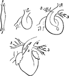
Fetal heart in successive stages of development. 1, venous extremity; 2, arterial extremity; 3, pulmonary branches; 4, ductus arteriosus.

Fetal Heart in Successive Stages of Development

Fetal heart in successive stages of development. 1, venous extremity; 2, arterial extremity; 3, pulmonary…

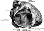
The right auricle of a fetal heart (eight months).

Fetal Heart

The right auricle of a fetal heart (eight months).

"One of the numberless microscopic bodies contained in semen, to which the seminal fluid owes its vitality, and which are the immediate and active means of impregnating or fertilizing the ovum of the female... I. broadest view; II. profile, of k, kernel or nucleus of the head, and m, filamentous body, ending in s, the long slender tail." —Whitney, 1889

Human Spermatozoa

"One of the numberless microscopic bodies contained in semen, to which the seminal fluid owes its vitality,…

Three stages in the development of the insula and the insular opercula.

Development of the Insula

Three stages in the development of the insula and the insular opercula.

Membranous labyrinth of a five months' fetus, viewed from its postero-mesial aspect.

Labyrinth of Fetus

Membranous labyrinth of a five months' fetus, viewed from its postero-mesial aspect.
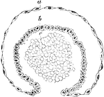
Transverse section of a developing Malpighian capsule and tuft from a fetus at about the fourth month. Labels: a, flattened cells growing to form the capsule; b, more rounded cells; continuos with the above, reflected around c, and finally enveloping it; c, mass of embryonic cells which later become developed into blood vessels.

Development of Malpighian Capsule

Transverse section of a developing Malpighian capsule and tuft from a fetus at about the fourth month.…
Three stages in the development of the medulla oblongata.

Development of Medulla Oblongata

Three stages in the development of the medulla oblongata.

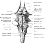
Back view of the medulla, pons, and mesencephalon of a full term human fetus.

Back View of Medulla, Pons, and Mesencephalon

Back view of the medulla, pons, and mesencephalon of a full term human fetus.
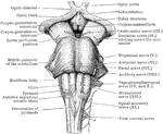
Front view of the medulla, pons, and mesencephalon of a full term human fetus.

Front view of Medulla, Pons, and Mesencephalon

Front view of the medulla, pons, and mesencephalon of a full term human fetus.
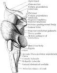
Lateral view of the medulla, pons, and mesencephalon of a full term human fetus.

Lateral View of Medulla, Pons, and Mesencephalon

Lateral view of the medulla, pons, and mesencephalon of a full term human fetus.

Transverse section through the closed part of the fetal medulla immediately above the decussation of the pyramids.

Transverse Section Through Closed Part of Fetal Medulla

Transverse section through the closed part of the fetal medulla immediately above the decussation of…

Transverse section through the lower end of the medulla of a full term fetus.

Transverse Section Through the the Medulla

Transverse section through the lower end of the medulla of a full term fetus.
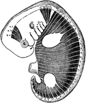
Scheme to illustrate the disposition of the myotomes in the embryo in relation to the head, trunk, and limbs. The muscles eventually form from these myotomes. Labels: A,B,C, First three cephalic myotomes. N,1,2,3,4, Last persisting cephalic myotomes. C,T,L,S,Co., The myotomes of the cervical, thoracic, lumbar, sacral and caudal regions. I.,II.,III.,IV.,V.,VI.,VII.,VIII.,IX.,X.,XI.,XII., Refer to the cranial nerves and the structures with which they may be embryologically associated.

Myotomes in an Embryo

Scheme to illustrate the disposition of the myotomes in the embryo in relation to the head, trunk, and…
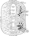
Diagram showing development of neural tube and crest.

Development of Neural Tube

Diagram showing development of neural tube and crest.

Transverse section through the early neural tube, diagrammatically represented. The left side of the section exhibits an earlier stage of development than the right side.

Section Through Early Neural Tube

Transverse section through the early neural tube, diagrammatically represented. The left side of the…
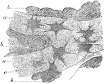
Osteoblasts from the parietal bone of a human embryo, thirteen weeks old. Labels: a, bony septa with cells of the lacunae; b, layers of osteoblasts; c, the latter in transition to bone corpuscles.

Osteoblasts

Osteoblasts from the parietal bone of a human embryo, thirteen weeks old. Labels: a, bony septa with…
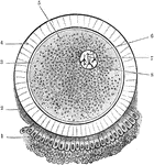
The ovum and its coverings. The corona radiata, which completely surrounds the ovum, is only represented in the lower part of the figure. Labels: 1, corona radiate; 2, granular layer; 3, vitelline membrane; 4, zona pellucida (oolemma); 5, vitellus or yolk; 6, germinal vesicle (nucleus); 7, germinal spot (nucleolus); 8, nuclear membrane.

Ovum

The ovum and its coverings. The corona radiata, which completely surrounds the ovum, is only represented…

Sectional views of the ovum, after segmentation.

Ovum After Segmentation

Sectional views of the ovum, after segmentation.

Diagram of a developing ovum, seen in longitudinal section. Labels: a, pericardium; b, bucco-pharyngeal; c, ectoderm; d, entoderm; e,placental area. f, spinal cord; i, brain; g, notochord; k, extra embryonic coelom; h, dorsal wall of alimentary canal.

Developing Ovum

Diagram of a developing ovum, seen in longitudinal section. Labels: a, pericardium; b, bucco-pharyngeal;…
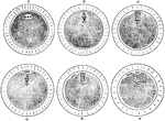
The maturation of the ovum. A, An ovum at the commencement process. B, After the formation of the spindle. The chromosomes are gathered at the equator of the spindle in groups of four, i.e. in tetrads, each which consists of two dyads. C, One apex of the spindle has projected into a bud on the surface, and the dyads have passed to the poles. D, The separation of the first polar body. E, The commencement of the second polar body; F, The completion of the second polar body.

Maturation of the Ovum

The maturation of the ovum. A, An ovum at the commencement process. B, After the formation of the spindle.…
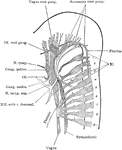
Reconstruction of peripheral nerves of human embryo of five weeks.

Peripheral Nerves of Human Embryo

Reconstruction of peripheral nerves of human embryo of five weeks.
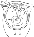
Diagram of an early stage of the formation of the human placenta. Labels: a, embryo; b, amnion; c, placental vessels; d, decidua reflexa; e, allantois; f, placental villi; g, mucous membrane.

Early Formation of the Placenta

Diagram of an early stage of the formation of the human placenta. Labels: a, embryo; b, amnion; c, placental…
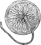
The organ of attachment of a vertebrate embryo or fetus to the wall of the uterus or womb of the female.

Human Placenta

The organ of attachment of a vertebrate embryo or fetus to the wall of the uterus or womb of the female.

Section of human placenta at end of pregnancy. The fetal blood vessels have been injected; the maternal blood spaces appear as clear areas surrounding the sections of the fetal villi.

Section of Placenta

Section of human placenta at end of pregnancy. The fetal blood vessels have been injected; the maternal…
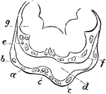
Extremities of a placental villus. Labels: a, lining membrane of the vascular system of the mother; b, cells immediately lining a; d, space between the maternal and fetal portion of the villus; e, internal membrane of the chorion; f, internal cells of the villus, or cells of the chorion; g, loop of umbilical vessels.

Placental Villus

Extremities of a placental villus. Labels: a, lining membrane of the vascular system of the mother;…

The developmental stages exhibited by a pyramidal cell of the brain. Labels: a, neuroblast with rudimentary axon, but no dendrites; b and c, the dendrites beginning to sprout out; d and e, further development of the dendrites and appearance of collateral branches on the axon.

Pyramidal Cells of the Brain

The developmental stages exhibited by a pyramidal cell of the brain. Labels: a, neuroblast with rudimentary…

The anal canal and rectum in a fetus. A, aged 4 to 5 months; B, 6 moths; C, 9 months. In each he anal canal is distinctly marked off from the rectum proper; the column of Morgagni and the rectal valve are distinct.

Rectum and Anal Canal in Fetus

The anal canal and rectum in a fetus. A, aged 4 to 5 months; B, 6 moths; C, 9 months. In each he anal…