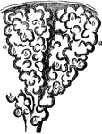
Two alveoli of the lung, highly magnified. Alveoli are cavities which are honeycombed with bulgings in order to increase the surface area exposed to air and covered by capillaries. Labels: b,b, bulgings of the alveoli, a, a.

Two Alveoli of the Lung

Two alveoli of the lung, highly magnified. Alveoli are cavities which are honeycombed with bulgings…
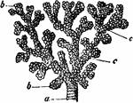
A small bronchial tube, a, dividing into its terminal branches, c; these have pouched or sacculated walls and end in the sacculated alveoli, b.

Bronchial Tube

A small bronchial tube, a, dividing into its terminal branches, c; these have pouched or sacculated…
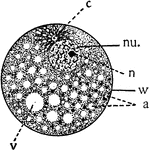
Diagram showing the principal parts of the cell and something of the protoplasmic architecture as it might appear while living. a, alveoli or spheres in the foam-work; c, centrosome; cy, cytoplasmic meshwork, containing granules; nu., nucleus; n, nucleolus; v. vacuole; w, cell wall.

Cell

Diagram showing the principal parts of the cell and something of the protoplasmic architecture as it…
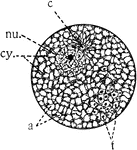
"Diagram showing the principal parts of the cell as it appeaers when killed and stained. The protoplasm shows more of a meshwork (cy), the spaces representing the alveoli. f, formed substance in alveoli." -Galloway, 1915

Cell Parts

"Diagram showing the principal parts of the cell as it appeaers when killed and stained. The protoplasm…
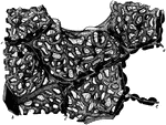
Section of lung with distended blood vessels, highly magnified. Labels: c,c, partitions between alveoli; b, small artery giving off capillaries to alveoli.

Section of Lung

Section of lung with distended blood vessels, highly magnified. Labels: c,c, partitions between alveoli;…

The lungs and air passages seen from the front. On the left of the figure the pulmonary tissue has been dissected away to show the ramifications of the bronchial tubes. Labels: a, larynx; b, trachea; d, right bronchus. The left bronchus is seen entering the root of its lung.

Lungs and Air Passages

The lungs and air passages seen from the front. On the left of the figure the pulmonary tissue has been…
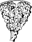
Structure of the lung. The lung has a serous coat; a sub-serous, elastic areolar tissue, investing the entire organ, and extending inward between the lobules; and the parenchyma, or true lung-tissue, composed of lobules and alveoli (or air-cells).

Structure of the Lungs

Structure of the lung. The lung has a serous coat; a sub-serous, elastic areolar tissue, investing the…
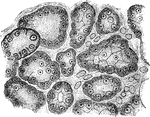
From the section through a mucous gland after prolonged electrical stimulation. The alveoli are lined with small granular cells.

Section of a Mucous Gland After Stimulation

From the section through a mucous gland after prolonged electrical stimulation. The alveoli are lined…
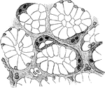
From the section through a mucous gland in a quiescent state. The alveoli are lined with transparent mucous cells, and outside these are the semilunes.

Section of a Mucous Gland

From the section through a mucous gland in a quiescent state. The alveoli are lined with transparent…
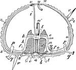
"Diagram of an Echinus (stripped of its spines). a, mouth; a', gullet; b, teeth; c, lips; d, alveoli; e, falces; f, f, auriculariae; g, retractor, and h, protractor, muscles of Aristotle's lantern; i, madreporic canal; k, circular ambulacral vessel; l, Polian pedicels; r, r, spines; s, tubercle; s', tubercle to which a spine is articulated; t, t, pedicellariae; u, anus; v, madreporic tubercle; x, ocular spot." -Whitney, 1911

Sea Urchin Section

"Diagram of an Echinus (stripped of its spines). a, mouth; a', gullet; b, teeth; c, lips; d, alveoli;…