Clipart tagged: ‘appendix’
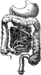
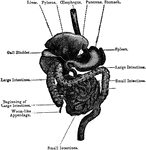
Alimentary Canal
Diagram of the abdominal part of the alimentary canal (digestive system). Labels: C, the cardiac, and…
Structure of the Appendix
Structure of the appendix. A. From a child two years old. B. From a male age 56. It will be observed…
Vermiform appendix
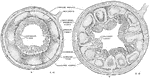
"A, a portion of the colon laid open to show the valve between the large and small intestine;…
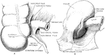
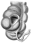
Caecal Folds and Fossae
The caecal folds and fossae. In A, the caecum is viewed from the front; the mesentery of the appendix…
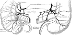
Blood Supply of Caecum and Appendix
The blood supply of the caecum and vermiform appendix. The illustration to the left gives a front view,…
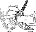
Arteries and Veins of the Cecum and Appendix
Arteries and veins of the cecum and vermiform appendix seen from behind.
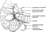
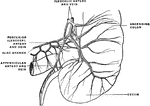
Arteries of the Cecum
Arteries of the cecum and of the appendix vermiformis and of the terminal portion of the ileum.