The ethmoid viewed from above.

Ethmoid Viewed from Above

The ethmoid viewed from above.

The ethmoid viewed from behind.

Ethmoid Viewed from Behind

The ethmoid viewed from behind.

The ethmoid viewed from right side.

Ethmoid Viewed from Side

The ethmoid viewed from right side.

Section showing nasal aspect of left lateral mass of ethmoid.

Section of the Ethmoid

Section showing nasal aspect of left lateral mass of ethmoid.

Shown is the base of the skull seen from above. Labels: 1, frontal bone; 2, slit for nasal nerve; 3, anterior ethmoidal foramen; 4, posterior ethmoidal foramen; 5, optic foramen; 6, foramen for internal carotid artery formed by anterior and middle clinoid processes; 7, lesser wing of sphenoid; 8, anterior clinoid process, in this case united on its inner side to the middle clinoid process; 9, posterior clinoid process; 10, foramen ovale; 11, groove for middle meningeal artery; 12, foramen spinosum; 13, Hiatus Fallopii; 14, line of petro-squamosal suture; 15, internal auditory meatus; 16, groove for superior petrosal sinus; 17, groove for sigmoid part of lateral sinus; 18, jugular foramen; 19, anterior condylic foramen; 20, groove for lateral sinus; 21, internal occipital protuberance; 22, ridge for attachment of falx cerebri; 23, fossa for the lodgment of the occipital lobe of the cerebrum; 24, ridge for attachment of the falx cerebelli; 25, fossa for the lodgment of the left cerebellar hemisphere; 26, foramen magnum; 27, groove for the sigmoid sinus turning into the jugular foramen; 28, groove for the inferior petrosal sinus running along the line of the suture between the petrous temporal and the basioccipital; 29, depression for the Gasserian ganglion; 30, middle cranial fossa for lodgment of temporal lobe of cerebrum; 31, foramen lacerum medium; 32, carotid groove; 33, dorsum sellae of sphenoid; 34, leads into foramen rotundum; 35, pituitary fossa; 36, olivary eminence of sphenoid; 37, anterior cranial fossa for lodgment of frontal lobes of cerebrum; 38, cribriform plate of ethmoid; 39, crista galli of ethmoid; 40, foramen caecum; 41, crest for attachment of falx cerebri.

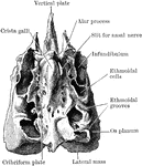
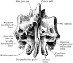
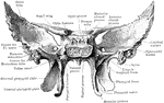
Base of the Skull Seen From Above

Shown is the base of the skull seen from above. Labels: 1, frontal bone; 2, slit for nasal nerve; 3,…

Shown is the inner aspect of the left half of the skull sagittally divided. Labels: 1, suture between parietal and temporal bone; 2, remains of subarcuate fossa; 3, grooves for branches of the middle meningeal artery; 4, dorsum sellae; 5, pituitary fossa; 6, anterior clinoid fossa; 7, optic foramen; 8, sphenoidal suture; 9, nasal surface of superior turbinated bone; 10, cribriform plate of ethmoid; 11, nasal surface of middle turbinated bone; 12, frontal sinus; 13, nasal spine; 14, nasal bone; 15, nasal process of superior maxilla; 16, middle meatus of nose; 17, directed towards opening of antrum; 18, nasal surface of inferior turbinated bone; 19, inferior meatus of nose; 20, anterior nasal spine; 21, anterior palatine canal; 22, palatal process of superior maxilla; 23, palatal process of palate bone; 24, posterior nasal spine; 25, hamular process of internal pterygoid plate; 26, external pterygoid plate; 27, superior meatus of nose; 28,sphenopalatine foramen; 29, pterygospinous ligament almost completely ossified to enclose a foramen; 30, styloid process of temporal bone; 31, alar spine of sphenoid; 32, mastoid process; 33, basion (midpoint of posterior border of foramen magnum); 34, internal auditory meatus; 35, anterior condylic foramen; 36, leading into jugular foramen; 37, opisthion (midpoint of posterior border of foramen magnum); 38, groove for sigmoid sinus; 39, opening of mastoid foramen; 40, for lateral sinus and attachment of tentorium cerebelli; 41, fossa for lodgment of cerebellar hemisphere; 42, internal occipital protuberance.

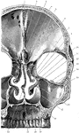
Skull Seen From Side

Shown is the inner aspect of the left half of the skull sagittally divided. Labels: 1, suture between…
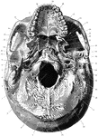
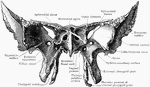
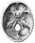
Shown is norma basalis, which refers to the base of the cranium. Labels: 1, external occipital crest; 2, superior curved line of the occipital bone; 3, foramen magnum; 4, occipital condyle; 5, digastric groove; 6, mastoid process; 7, external auditory meatus; 8, styloid process; 9, glenoid fossa; 10, foramen spinosum; 11, sphenoidal spine; 12, foramen ovale; 13, external pterygoid plate; 14, hamular process of internal pterygoid plate; 15, nasal septum; 16, posterior nasal spine; 17, horizontal plate of palate bone; 18, palatal process of superior maxilla; 19, anterior palatine canal; 20, intermaxillary suture; 21, posterior palatine canal; 22, malar process of superior maxilla; 23, spheno-maxillary fissure; 24, zygomatic fossa; 25, zygomatic arch; 26, posterior openings of left nasal fossa; 27, pterygoid fossa; 28, scaphoid fossa; 29, foramen lacerum medium; 30, opening of osseous Eustachian canal; 31, carotid canal; 32, jugular fossa; 33, stylo-mastoid foramen; 34, jugular process of the occipital bone; 35, groove for occipital artery; 36, mastoid foramen; 37, posterior condylic foramen; 38, inferior curved line of occipital bone; 39, external occipital protuberance.

Base of the Skull

Shown is norma basalis, which refers to the base of the cranium. Labels: 1, external occipital crest;…

Shown is a coronal section passing inferiorly through interval between between the first and second molar teeth. Labels: 1, groove of superior longitudinal sinus; 2,crest for attachment of falx cerebri; 3, crista galli of ethmoid; 4, cristiform plate of ethmoid; 5, perpendicular plate of ethmoid, assisting in the formation of nasal septum; 6, lateral mass of ethmoid consisting of the ethmoidal cells; 7, os planum of ethmoid; 8, middle meatus of nose; 9, middle turbinated bone; 10, opening from the middle meatus; 11, orbital plate of superior maxilla; 12, frontomalar suture; 13, infraorbital groove; 14, antrum or maxillary sinus; 15, canal for anterior dental nerve and vessels exposed; 16, inferior meatus of nose; 17, alveolar process of the superior maxilla; 18, nasal surface of inferior turbinated bone; 19, groove for anterior palatine nerve and vessels; 20, palatal process of superior maxilla; 21, maxillary crest forming part of nasal septum; 22, vomer forming part of nasal septum.

Coronal Section of Skull

Shown is a coronal section passing inferiorly through interval between between the first and second…
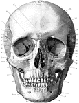
Shown is norma frontalis, which refers to the front of the skull. Labels: 1, mental protuberance; 2, body of lower jaw; 3, ramus of lower jaw; 4, anterior nasal spine; 5, canine fossa; 6, infraorbital canal; 7, malar canal; 8 orbital surface of superior maxilla; 9, temporal fossa; 10, os planum of ethmoid; 11, sphenoidal fissure; 12, lachrymal bone and groove; 13, optic foramen; 14, orbital foramina; 15, temporal ridge; 16, supraorbital notch; 17, glabella; 18, frontal eminence; 19, superciliary ridge; 20, parietal bone; 21, front-nasal suture; 22, pterion; 23, great wing of sphenoid; 24, orbital surface of great wing of sphenoid; 25, squamous temporal; 26, left nasal bone; 27, malar bone; 28, sphenomaxillary fissure; 29, zygomatic arch; 30, anterior nasal aperture, displaying nasal septum and inferior and middle turbinated bones; 31, mastoid process; 32, incisor fossa; 33, angle of jaw; 34, mental foramen; 35, symphysis.

Front of the Skull

Shown is norma frontalis, which refers to the front of the skull. Labels: 1, mental protuberance; 2,…
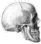
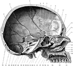
Shown is norma lateralis, which refers to the side of the skull. Labels: 1, mental foramen; 2, body of lower jaw; 3, superior maxillary; 4, ramus of lower jaw; 5, zygomatic arch; 6, styloid process; 7, external auditory meatus; 8, mastoid process; 9, asterion; 10, superior curved line of occipital bone; 11, external occipital protuberance; 12, lambdoid suture; 13, occipital bone; 14, lambda; 15, obelion placed between the two parietal foramina; 16, parietal bone; 17, lower temporal ridge; 18, upper temporal ridge; 19, squamous part of temporal bone; 20, bregma; 21, coronal suture; 22, stephanion; 23, frontal bone; 24, pterion; 25, temporal fossa; 26, great wing of sphenoid; 27, malar bone; 28, malar canal; 29, lachrymal bone; 30, nasal bone; 31, infraorbital canal; 32, anterior nasal aperture.

Side of the Skull

Shown is norma lateralis, which refers to the side of the skull. Labels: 1, mental foramen; 2, body…

The sphenoid as seen from behind.

Sphenoid Viewed from Behind

The sphenoid as seen from behind.

The sphenoid as seen from the front.

Sphenoid Viewed from the Front

The sphenoid as seen from the front.